Korean J Neurotrauma.
2019 Oct;15(2):182-186. 10.13004/kjnt.2019.15.e20.
Spontaneous Intracranial and Spinal Subdural Hematoma: A Case Report
- Affiliations
-
- 1Department of Neurosurgery, College of Medicine, Chosun University, Gwangju, Korea. ns64902@hanmail.net
- 2Department of Radiology, College of Medicine, Chosun University, Gwangju, Korea.
- KMID: 2461125
- DOI: http://doi.org/10.13004/kjnt.2019.15.e20
Abstract
- Spinal subdural hematoma (SDH) is rarely reported, and their simultaneous occurrence with intracranial SDH is even more rare. A 67-year-old male patient with a history of posterolateral fusion to treat an L2 burst fracture came to our outpatient clinic due to an inability to walk by himself over the previous 3 weeks. A neurological examination revealed that the patient was alert with occasional confusion and slight motor weakness in the lower extremities. Brain and lumbar spine magnetic resonance imaging (MRI) was then performed. A brain MRI revealed a large subacute SDH along the right cerebral convexity and falx cerebri with midline shifting, and a spine MRI revealed a right side-predominant subacute SDH extending from L4 to S1. For treatment, burr hole trephination of the intracranial SDH and fluoroscopy-guided lumbar puncture of the spinal SDH were performed and resulted in a favorable outcome. This is a report of a rare case of spontaneous intracranial and lumbar spine SDH. We include a review of the current literature and a discussion of the pathogenesis of this condition in this report.
Keyword
MeSH Terms
Figure
-
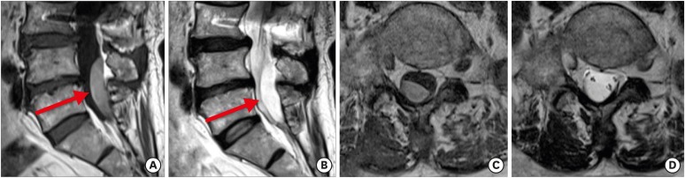
FIGURE 1 Lumbar spine MRI images of the patient. Sagittal T1-weighted (A) and T2-weighted (B) images reveal a hematoma (arrows) in the dorsal spine at the L4–S1 level. Axial T1-weighted (C) and T2-weighted (D) images at the L5 level reveal a subacute subdural hematoma. The hematoma is right-side dominant.MRI: magnetic resonance imaging.
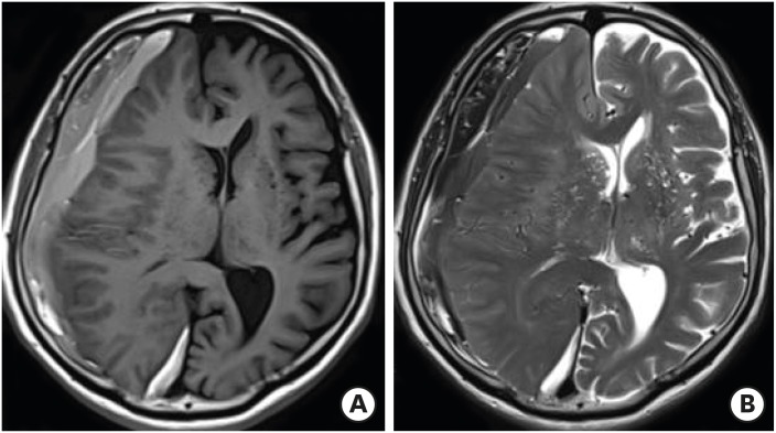
FIGURE 2 Brain MRI images of the patient. Axial T1-weighted (A) and T2-weighted (B) MRI images demonstrate a large right-side subdural hematoma with slight hyperintensity compared to CSF. This MRI finding is typical of subacute SDH.MRI: magnetic resonance imaging, CSF: cerebrospinal fluid, SDH: spinal subdural hematoma.
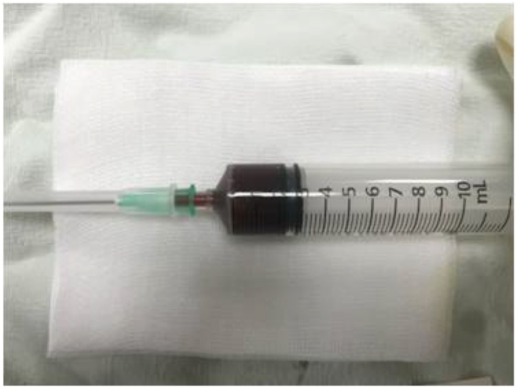
FIGURE 3 Dark-brownish blood obtained with fluoroscopy-guided lumbar puncture.
Reference
-
1. Domenicucci M, Ramieri A, Paolini S, Russo N, Occhiogrosso G, Di Biasi C, et al. Spinal subarachnoid hematomas: our experience and literature review. Acta Neurochir (Wien). 2005; 147:741–750. PMID: 15711890.
Article2. Kanamaru H, Kanamaru K, Araki T, Hamada K. Simultaneous spinal and intracranial chronic subdural hematoma cured by craniotomy and laminectomy: a video case report. Case Rep Neurol. 2016; 8:72–77. PMID: 27194987.
Article3. Kim MS, Sim SY. Spinal subdural hematoma associated with intracranial subdural hematoma. J Korean Neurosurg Soc. 2015; 58:397–400. PMID: 26587198.
Article4. Kim YW, Hwang SO, Kang HS, Cha KC. The gradient between arterial and end-tidal carbon dioxide predicts in-hospital mortality in post-cardiac arrest patient. Am J Emerg Med. 2019; 37:1–4. PMID: 29685358.
Article5. Kokubo R, Kim K, Mishina M, Isu T, Kobayashi S, Yoshida D, et al. Prospective assessment of concomitant lumbar and chronic subdural hematoma: is migration from the intracranial space involved in their manifestation? J Neurosurg Spine. 2014; 20:157–163. PMID: 24286531.
Article6. Kwon OI, Son DW, Kim YH, Kim YS, Sung SK, Lee SW, et al. Migration of an intracranial subdural hematoma to the spinal subdural space: a case report. Korean J Spine. 2015; 12:207–209. PMID: 26512286.
Article7. Lecouvet FE, Annet L, Duprez TP, Cosnard G, Scordidis V, Malghem J. Uncommon magnetic resonance imaging observation of lumbar subdural hematoma with cranial origin. J Comput Assist Tomogr. 2003; 27:530–533. PMID: 12886137.
Article8. Lin JC, Layman K. Spontaneous spinal subdural hematoma of intracranial origin presenting as back pain. J Emerg Med. 2014; 47:552–556. PMID: 25216539.
Article9. Matsumoto H, Matsumoto S, Yoshida Y. Concomitant intracranial chronic subdural hematoma and spinal subdural hematoma: a case report and literature review. World Neurosurg. 2016; 90:706.e1–706.e9.
Article10. Sánchez-Menoyo JL, Ruiz-Ojeda J, Martínez-Arroyo A, García-Moncó JC, Aduna-De Paz M, Vicente-Olabarría I. Spinal cord hemorrhage complicating diagnostic lumbar puncture. Rev Neurol. 2009; 48:418–420. PMID: 19340782.11. Satyarthee GD, Ahmad F. Spontaneous concurrent intraspinal and intracranial subdural hematoma: management and review of literature. J Pediatr Neurosci. 2018; 13:24–27. PMID: 29899767.12. Seung WB, Jeong JH. Postoperative subarachnoid hemorrhage and multipunctate intracerebral hemorrhages following evacuation of bilateral chronic subdural hematomas. Korean J Neurotrauma. 2017; 13:149–152. PMID: 29201851.
Article13. Singh DK, Chauhan M, Gupta V, Chopra S, Bagaria HR. Spinal subdural hematoma: a rare complication of spinal anesthesia: a case report. Turk Neurosurg. 2008; 18:324–326. PMID: 18814128.14. Yamaguchi S, Kurisu K, Arita K, Takeda M, Tani I, Araki O. Simultaneous cranial and spinal subdural hematoma. Neurol Med Chir (Tokyo). 2005; 45:645–649. PMID: 16377954.
- Full Text Links
-
- Actions
-
Cited
- CITED
-
- Close
- Share
-
- Similar articles
-
- Spontaneous Spinal Subdural and Subarachnoid Hemorrhage with Concomitant Intracerebral Hemorrhage: A Case Report
- Spontaneous Concomitant Intracranial and Spinal Subdural Hematomas in Association with Anticoagulation Therapy
- Simultaneous Intracranial and Spinal Subdural Hematoma: Two Case Reports
- Traumatic Spinal Subdural Hematoma Accompanying intracranial hematoma: Spontaneous Resolution after Pumbar Puncture
- Intraoperative Development of Contralateral Subdural Hematoma during Evacuation of Acute Subdural Hematoma: Case Report
