Screening of Sera from Patients with Pancreatitis by an Apoptosis Assay of Skin-derived Cells
- Affiliations
-
- 1Department of Senior Healthcare, BK21 Plus Program, Graduate School, Eulji University, Daejeon, Korea. kanghg@eulji.ac.kr
- 2Division of Gastroenterology, Department of Internal Medicine, Eulji University Eulji Hospital, Seoul, Korea.
- 3Department of Biomedical Laboratory Science, College of Health Sciences, Eulji University, Seongnam, Korea.
- 4Department of Laboratory Medicine, Seongnam Central Hospital, Seongnam, Korea.
- KMID: 2460987
- DOI: http://doi.org/10.4166/kjg.2019.74.4.219
Abstract
- BACKGROUND/AIMS
An excessive inflammatory response is typical in acute pancreatitis and a significant cause of early mortality in severe acute pancreatitis. This is believed to be caused by inflammatory molecules or upregulated cytokine levels in the serum of patients. The aim of this study was to identify the serum-mediated apoptosis-inducing effects in acute pancreatitis patients.
METHODS
A skin tissue-derived cell line, BJ, was treated for 24 hours with the sera of 22 healthy volunteers (control) and 71 acute pancreatitis patients (22 with gallstone pancreatitis, 16 with alcoholic pancreatitis, and 11 with pancreatitis with other causes) collected at the time of hospital admission (active) and discharge (resolved). Apoptosis was analyzed by flow cytometry.
RESULTS
The average percentage of living cells, early apoptotic cells, and late apoptotic cells ranged from 78.8% to 85.0%, 5.5% to 7.3%, and 7.7% to 13.1%, respectively. The number of live cells increased significantly using the serum from the resolved state of gallstone-induced pancreatitis. In addition, the number of early apoptotic cells increased significantly using the serum from the resolved state of pancreatitis with other causes. The number of late apoptotic cells decreased significantly with the serum from the resolved state compared to the active state of gallstone- and alcohol-induced pancreatitis.
CONCLUSIONS
Serum samples from patients with pancreatitis induced a change in the apoptosis profiles of skin-derived cells. These results indicate changes in the serum components in patients with acute pancreatitis.
MeSH Terms
Figure
-
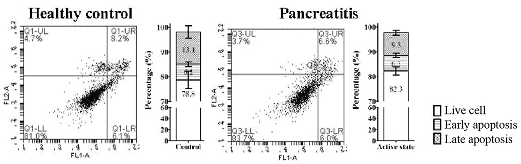
Fig. 1 Apoptosis analysis of sera from healthy control and acute pancreatitis patients of BJ cell lines. Scatter plot of Annexin V and propidium iodide staining for healthy control serum- and patient serum-treated cells. Quantitative analysis of the percentage of live cells, and the early and late apoptotic cells by flow cytometry are shown. Q, quadrant; UL, upper left; UR, upper right; LL, lower left; LR, lower right.
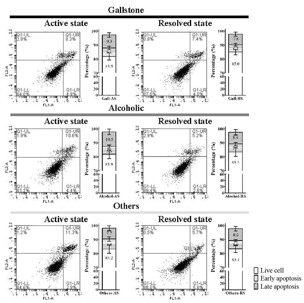
Fig. 2 Flow cytometry analysis of sera from patients with acute pancreatitis includes apoptosis of BJ cell lines. Scatter plot of Annexin V and propidium iodide staining acute pancreatitis patient serum-treated cells. Quantitative analysis of the percentage of live cells, and early and late apoptotic cells by flow cytometry analysis are shown. Error bars, mean±standard error. AS, active state; RS, resolved state; Q, quadrant; UL, upper left; UR, upper right; LL, lower left; LR, lower right.
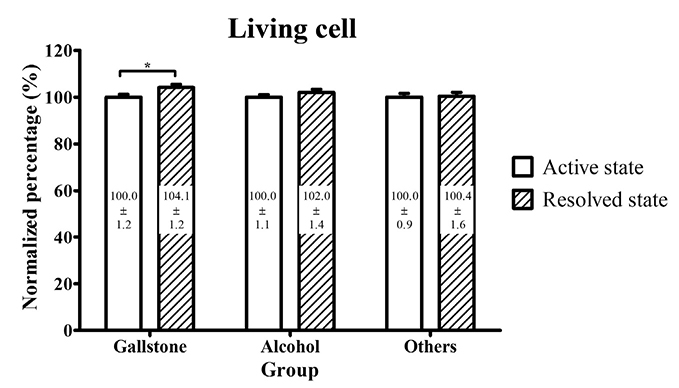
Fig. 3 Comparative analysis of living cells in an apoptosis assay. Error bars, mean±standard error. *p-value <0.05.
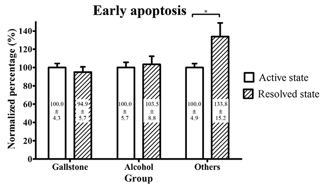
Fig. 4 Comparative analysis of early apoptotic cells in an apoptosis assay. Error bars, mean±standard error. *p-value <0.05.

Fig. 5 Comparative analysis of late apoptotic cells in an apoptosis assay. Error bars, mean±standard error. *p-value <0.05.
Reference
-
1. Medzhitov R. Origin and physiological roles of inflammation. Nature. 2008; 454:428–435.
Article2. Gottschalk TA, Tsantikos E, Hibbs ML. Pathogenic inflammation and its therapeutic targeting in systemic lupus erythematosus. Front Immunol. 2015; 6:550.
Article3. Fahy JV. Type 2 inflammation in asthma--present in most, absent in many. Nat Rev Immunol. 2015; 15:57–65.4. Minasyan H. Sepsis and septic shock: pathogenesis and treatment perspectives. J Crit Care. 2017; 40:229–242.
Article5. Dumnicka P, Maduzia D, Ceranowicz P, Olszanecki R, Drożdż R, Kuśnierz-Cabala B. The interplay between inflammation, coagulation and endothelial injury in the early phase of acute pancreatitis: clinical implications. Int J Mol Sci. 2017; 18:E354.
Article6. Beger HG, Rau B, Mayer J, Pralle U. Natural course of acute pancreatitis. World J Surg. 1997; 21:130–135.
Article7. Meier RF, Sobotka L. Basics in clinical nutrition: nutritional support in acute and chronic pancreatitis. E Spen Eur E J Clin Nutr Metab. 2010; 5:e58–e62.
Article8. Appelros S, Borgström A. Incidence, aetiology and mortality rate of acute pancreatitis over 10 years in a defined urban population in Sweden. Br J Surg. 1999; 86:465–470.
Article9. Harrison DA, D'amico G, Singer M. The Pancreatitis Outcome Prediction (POP) score: a new prognostic index for patients with severe acute pancreatitis. Crit Care Med. 2007; 35:1703–1708.
Article10. Vege SS, Gardner TB, Chari ST, et al. Low mortality and high morbidity in severe acute pancreatitis without organ failure: a case for revising the Atlanta classification to include "moderately severe acute pancreatitis". Am J Gastroenterol. 2009; 104:710–715.
Article11. Banks PA, Bollen TL, Dervenis C, et al. Classification of acute pancreatitis--2012: revision of the Atlanta classification and definitions by international consensus. Gut. 2013; 62:102–111.
Article12. Kang R, Lotze MT, Zeh HJ, Billiar TR, Tang D. Cell death and DAMPs in acute pancreatitis. Mol Med. 2014; 20:466–477.
Article13. Wang G, Han B, Zhou H, et al. Inhibition of hydrogen sulfide synthesis provides protection for severe acute pancreatitis rats via apoptosis pathway. Apoptosis. 2013; 18:28–42.
Article14. Hasan SI, Mohd Ashari NS, Mohd Daud K, Che Husin CM. High occurrence of in vitro apoptosis of lymphocytes induced by serum from systemic lupus erythematosus patients is associated with increased serum levels of anti-C1q autoantibodies. Int J Rheum Dis. 2013; 16:430–436.15. Feltham R, Vince JE, Lawlor KE. Caspase-8: not so silently deadly. Clin Transl Immunology. 2017; 6:e124.
Article16. Jorgensen I, Miao EA. Pyroptotic cell death defends against intracellular pathogens. Immunol Rev. 2015; 265:130–142.
Article17. Kolb JP, Oguin TH 3rd, Oberst A, Martinez J. Programmed cell death and inflammation: winter is coming. Trends Immunol. 2017; 38:705–718.
Article18. Aguilar-Lemarroy A, Romero-Ramos JE, Olimon-Andalon V, et al. Apoptosis induction in Jurkat cells and sCD95 levels in women's sera are related with the risk of developing cervical cancer. BMC Cancer. 2008; 8:99.
Article19. Makino N, Maeda T, Sugano M, Satoh S, Watanabe R, Abe N. High serum TNF-α level in type 2 diabetic patients with microangiopathy is associated with eNOS down-regulation and apoptosis in endothelial cells. J Diabetes Complications. 2005; 19:347–355.
Article20. Gukovskaya AS, Gukovsky I, Zaninovic V, et al. Pancreatic acinar cells produce, release, and respond to tumor necrosis factor-alpha. Role in regulating cell death and pancreatitis. J Clin Invest. 1997; 100:1853–1862.
Article21. Norman JG, Fink GW, Franz MG. Acute pancreatitis induces intrapancreatic tumor necrosis factor gene expression. Arch Surg. 1995; 130:966–970.
Article22. Gu H, Werner J, Bergmann F, Whitcomb DC, Büchler MW, Fortunato F. Necro-inflammatory response of pancreatic acinar cells in the pathogenesis of acute alcoholic pancreatitis. Cell Death Dis. 2013; 4:e816.
Article23. Montecucco F, Mach F, Lenglet S, et al. Treatment with Evasin-3 abrogates neutrophil-mediated inflammation in mouse acute pancreatitis. Eur J Clin Invest. 2014; 44:940–950.
Article24. Folias AE, Penaranda C, Su AL, Bluestone JA, Hebrok M. Aberrant innate immune activation following tissue injury impairs pancreatic regeneration. PLoS One. 2014; 9:e102125.
Article
- Full Text Links
-
- Actions
-
Cited
- CITED
-
- Close
- Share
-
- Similar articles
-
- The Effect of Pemphigus Sera on Apoptosis of Keratinocytes
- Cerulein Pancreatitis: Oxidative Stress, Inflammation, and Apoptosis
- MSCs-Derived miR-150-5p-Expressing Exosomes Promote Skin Wound Healing by Activating PI3K/AKT Pathway through PTEN
- Usefulness of actim Pancreatitis(R) Test for Screening of Acute Pancreatitis in The ED
- Procurement of HLA Class I Antisera from Multiparous Blood Donors
