J Korean Ophthalmol Soc.
2019 Sep;60(9):874-880. 10.3341/jkos.2019.60.9.874.
Retinal Nerve Fiber Layer and Macular Retinal Ganglion Cell Layer Thicknesses in Healthy Korean Children
- Affiliations
-
- 1Myung-Gok Eye Research Institute, Kim's Eye Hospital, Konyang University College of Medicine, Seoul, Korea. brainh@hanmail.net
- KMID: 2459132
- DOI: http://doi.org/10.3341/jkos.2019.60.9.874
Abstract
- PURPOSE
To measure the thicknesses of the circumpapillary retinal nerve fiber layer (RNFL) and macular retinal ganglion cell-inner plexiform layer (GCIPL) by optical coherence tomography (OCT) in healthy Korean children and adolescents.
METHODS
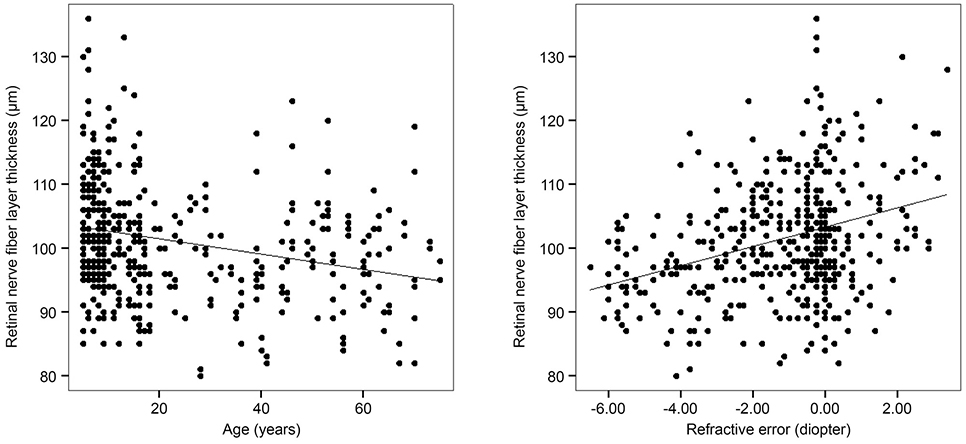
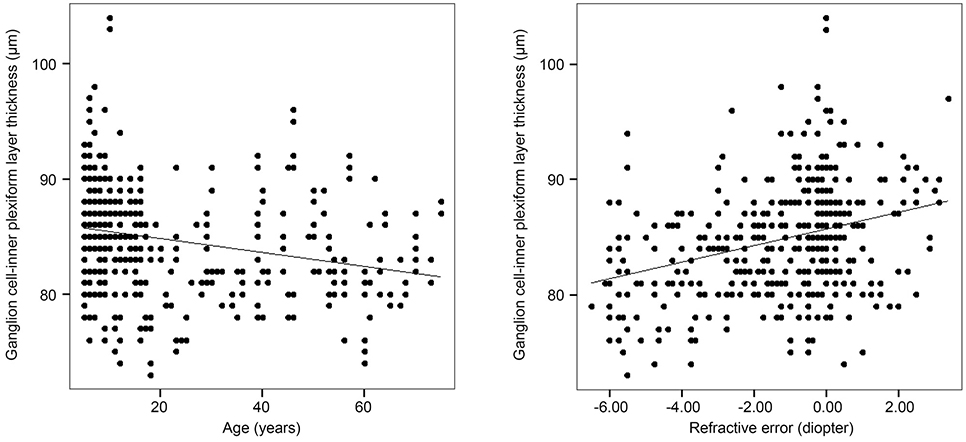
Circumpapillary RNFL and macular GCIPL thicknesses were measured by OCT in 352 healthy eyes of 352 children and adolescents (child-adolescent group) aged 5-17 years and in 159 healthy eyes of 159 adults (adult group) aged 18-75 years. The difference in RNFL and GCIPL thicknesses between the groups and the effects of age and refractive errors on the measurements were evaluated.
RESULTS
The mean age of the children and adults were 9.7 ± 3.6 and 44.7 ± 15.7 years, respectively. The mean of the average RNFL and GCIPL thicknesses were 103.1 ± 9.2 and 85.7 ± 4.6 µm, respectively, in the children group and 97.8 ± 8.2 and 82.9 ± 4.4 µm, respectively, in the adult group. The child-adolescent group had greater RNFL and GCIPL thicknesses compared to the adult group in all areas (p < 0.05) with the exception of the RNFL thickness in the temporal quadrant (p = 0.555). A thinner RNFL and thinner GCIPL were significantly associated with older age and greater myopia (p < 0.001).
CONCLUSIONS
The mean RNFL and GCIPL thicknesses measured by OCT in the healthy eyes of children and adolescents were 103.1 and 85.7 µm, respectively. Children and adolescents had a thicker RNFL and GCIPL compared to adults.
Keyword
MeSH Terms
Figure
Reference
-
1. Gospe SM, Bhatti MT, El-Dairi MA. Emerging applications of optical coherence tomography in pediatric optic neuropathies. Semin Pediatr Neurol. 2017; 24:135–142.
Article2. Lee H, Proudlock FA, Gottlob I. Pediatric optical coherence tomography in clinical practice-recent progress. Invest Ophthalmol Vis Sci. 2016; 57:OCT69–OCT79.
Article3. Srinivasan S, Addepalli UK, Rao HL, et al. Spectral domain optical coherence tomography in children operated for primary congenital glaucoma. Br J Ophthalmol. 2014; 98:162–165.
Article4. Barrio-Barrio J, Noval S, Galdós M, et al. Multicenter Spanish study of spectral-domain optical coherence tomography in normal children. Acta Ophthalmol. 2013; 91:e56–e63.
Article5. Elía N, Pueyo V, Altemir I, et al. Normal reference ranges of optical coherence tomography parameters in childhood. Br J Ophthalmol. 2012; 96:665–670.
Article6. Rao A, Sahoo B, Kumar M, et al. Retinal nerve fiber layer thickness in children <18 years by spectral-domain optical coherence tomography. Semin Ophthalmol. 2013; 28:97–102.7. Goh JP, Koh V, Chan YH, Ngo C. Macular ganglion cell and retinal nerve fiber layer thickness in children with refractive errors-an optical coherence tomography study. J Glaucoma. 2017; 26:619–625.
Article8. Turk A, Ceylan OM, Arici C, et al. Evaluation of the nerve fiber layer and macula in the eyes of healthy children using spectral-domain optical coherence tomography. Am J Ophthalmol. 2012; 153:552–559.e1.
Article9. Yanni SE, Wang J, Cheng CS, et al. Normative reference ranges for the retinal nerve fiber layer, macula, and retinal layer thicknesses in children. Am J Ophthalmol. 2013; 155:354–360.e1.
Article10. Tsai DC, Huang N, Hwu JJ, et al. Estimating retinal nerve fiber layer thickness in normal schoolchildren with spectral-domain optical coherence tomography. Jpn J Ophthalmol. 2012; 56:362–370.
Article11. Totan Y, GüraXMLLink_XYZaç FB, Güler E. Evaluation of the retinal ganglion cell layer thickness in healthy Turkish children. J Glaucoma. 2015; 24:e103–e108.
Article12. Park JM, Choi YJ, Kim DH. The analysis of peripapillary RNFL, macula and macular ganglion cell layer thickness in patients with monocular amblyopia using SD-OCT. J Korean Ophthalmol Soc. 2016; 57:98–105.
Article13. Lee YP, Ju YS, Choi DG. Ganglion cell-inner plexiform layer thickness by swept-source optical coherence tomography in healthy Korean children: normative data and biometric correlations. Sci Rep. 2018; 8:10605.
Article14. Yoo YJ, Hwang JM, Yang HK. Inner macular layer thickness by spectral domain optical coherence tomography in children and adults: a hospital-based study. Br J Ophthalmol. 2019; 01. 12. DOI: 10.1136/bjophthalmol-2018-312349. [Epub ahead of print].
Article15. Hong S, Kim SM, Park K, et al. Adjusted color probability codes for peripapillary retinal nerve fiber layer thickness in healthy Koreans. BMC Ophthalmol. 2014; 14:38.
Article16. Lee JY, Hwang YH, Lee SM, Kim YY. Age and retinal nerve fiber layer thickness measured by spectral domain optical coherence tomography. Korean J Ophthalmol. 2012; 26:163–168.
Article17. Kim EJ, Hong S, Kim CY, et al. Attenuated age-related thinning of peripapillary retinal nerve fiber layer in long eyes. Korean J Ophthalmol. 2011; 25:248–251.
Article18. Mwanza JC, Durbin MK, Budenz DL;. Cirrus OCT Normative Database Study Group. Interocular symmetry in peripapillary retinal nerve fiber layer thickness measured with the Cirrus HD-OCT in healthy eyes. Am J Ophthalmol. 2011; 151:514–521.e1.
Article19. Knight OJ, Girkin CA, Budenz DL, et al. Effect of race, age, and axial length on optic nerve head parameters and retinal nerve fiber layer thickness measured by Cirrus HD-OCT. Arch Ophthalmol. 2012; 130:312–318.
Article20. Lee YG, Hwang YH. A comparison of retinal nerve fiber layer thickness measured using five different optical coherence tomography devices. J Korean Ophthalmol Soc. 2018; 59:261–267.
Article21. Samarawickrama C, Wang JJ, Huynh SC, et al. Ethnic differences in optic nerve head and retinal nerve fibre layer thickness parameters in children. Br J Ophthalmol. 2010; 94:871–876.
Article22. Girkin CA, McGwin G Jr, Sinai MJ, et al. Variation in optic nerve and macular structure with age and race with spectral-domain optical coherence tomography. Ophthalmology. 2011; 118:2403–2408.
Article23. Leung CK, Yu M, Weinreb RN, et al. Retinal nerve fiber layer imaging with spectral-domain optical coherence tomography: a prospective analysis of age-related loss. Ophthalmology. 2012; 119:731–737.24. Hwang YH, Yoo C, Kim YY. Myopic optic disc tilt and the characteristics of peripapillary retinal nerve fiber layer thickness measured by spectral-domain optical coherence tomography. J Glaucoma. 2012; 21:260–265.
Article25. Kang SH, Hong SW, Im SK, et al. Effect of myopia on the thickness of the retinal nerve fiber layer measured by Cirrus HD optical coherence tomography. Invest Ophthalmol Vis Sci. 2010; 51:4075–4083.
Article26. Mwanza JC, Durbin MK, Budenz DL, et al. Profile and predictors of normal ganglion cell-inner plexiform layer thickness measured with frequency-domain optical coherence tomography. Invest Ophthalmol Vis Sci. 2011; 52:7872–7879.
Article27. Koh VT, Tham YC, Cheung CY, et al. Determinants of ganglion cell-inner plexiform layer thickness measured by high-definition optical coherence tomography. Invest Ophthalmol Vis Sci. 2012; 53:5853–5859.
Article
- Full Text Links
- Actions
-
Cited
- CITED
-
- Close
- Share
- Similar articles
-
- Comparison of OCT Parameters between the Dominant and Nondominant Eye
- The Normative Retinal and Choroidal Thicknesses of the Rabbit as Revealed by Spectral Domain Optical Coherence Tomography
- Retinal Nerve Fiber Layer and Macular Ganglion Cell Thicknesses in Situs Inversus of Optic Discs
- Thicknesses of the Fovea and Retinal Nerve Fiber Layer in Amblyopic and Normal Eyes in Children
- Thicknesses of Macular Retinal Layer and Peripapillary Retinal Nerve Fiber Layer in Patients with Hyperopic Anisometropic Amblyopia


