Dermoscopic Evolution of Pediatric Nevi
- Affiliations
-
- 1Department of Dermatoveneorology, Bezmialem Vakif University, Istanbul, Turkey. fpelinozgen@hotmail.com
- 2Bezmialem Vakif University Faculty of Medicine, Istanbul, Turkey.
- KMID: 2456228
- DOI: http://doi.org/10.5021/ad.2019.31.5.518
Abstract
- BACKGROUND
The incidence of pediatric melanoma is very rare. Dermoscopic features help to distinguish pediatric melanoma and common nevi.
OBJECTIVE
To study the evolution of dermoscopic findings in benign nevi in childhood through serial observation and photography.
METHODS
We examined 504 melanocytic lesions in 100 patients. From each participant, dermoscopic images of the nevi from 4-year dermoscopic follow-up were obtained, including randomly selected nevi.
RESULTS
The most common dermoscopic patterns were homogeneous (193 nevi; 38.3%), globular (92 nevi; 18.3%), and reticular (86 nevi; 17.1%). Dermoscopic pattern changes were detected in 27% of patients aged 2~10 years and in 20% of patients aged 11~16 years. The main pattern changes consisted of the transition from homogeneous to globular-homogeneous (16%), from homogeneous to reticular-homogeneous (12%) and from globular to globular-homogeneous (10%). Although 257 of the 504 nevi (51.0%) have stable duration without size changes, 169 of the 504 nevi (33.5%) were enlarged, and 78 of the 504 nevi (15.5%) had become smaller.
CONCLUSION
These results contrast with the prevailing view that dermoscopic patterns in pediatric nevi are usually characterized by globular patterns and that melanocytic nevi generally undergo a characteristic transition from a globular pattern to a reticular pattern. Fifty one percent of patients did not exhibit a size change. While 33% of patients had symmetrical enlargement, 15% of patients had involution. Therefore, enlargement is a common dermoscopic change in pediatric nevi, and is not a specific sign of pediatric melanoma.
Keyword
Figure
-
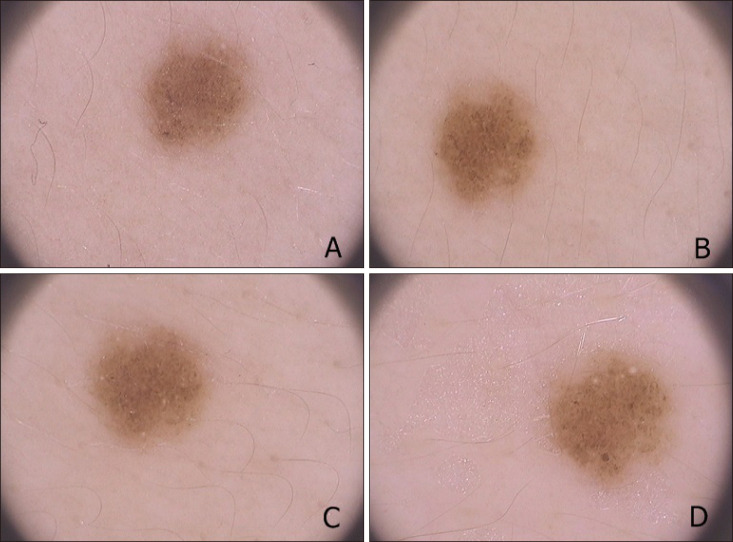
Fig. 1 A 9-year-old boy, nevus on the arm, dermoscopic change of homogeneous pattern to homogeneous-globular pattern (A: baseline, B: 12 months after, C: 24 months after, D: 48 months after).
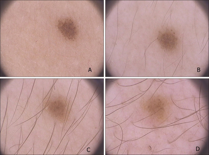
Fig. 2 A 14-year-old boy, nevus on the leg, dermoscopic change of globular pattern to homogeneous pattern (A: baseline, B: 12 months after, C: 24 months after, D: 48 months after).
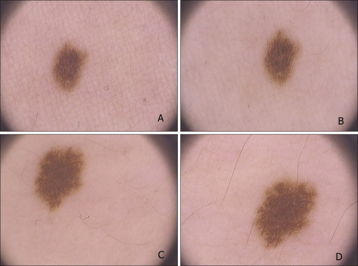
Fig. 3 A 10-year-old girl, nevus on the back, dermoscopic change of homogeneous pattern to reticular pattern, enlargement of nevus (A: baseline, B: 12 months after, C: 24 months after, D: 48 months after).
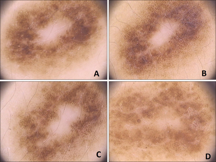
Fig. 4 A 12-year-old girl, nevus on the back, dermoscopic change of homogeneous-globular pattern to homogeneous-reticular pattern (A: baseline, B: 12 months after, C: 24 months after, D: 48 months after).
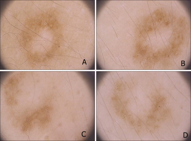
Fig. 5 A 8-year-old boy, nevus on the back, dermoscopy of homogeneous-reticular pattern, fading in color (A: baseline, B: 12 months after, C: 24 months after, D: 48 months after).
Reference
-
1. Fitzpatrick TB. The validity and practicality of sun-reactive skin types I through VI. Arch Dermatol. 1988; 124:869–871. PMID: 3377516.
Article2. Argenziano G, Soyer HP, Chimenti S, Talamini R, Corona R, Sera F, et al. Dermoscopy of pigmented skin lesions: results of a consensus meeting via the Internet. J Am Acad Dermatol. 2003; 48:679–693. PMID: 12734496.3. Kittler H, Pehamberger H, Wolff K, Binder M. Follow-up of melanocytic skin lesions with digital epiluminescence microscopy: patterns of modifications observed in early melanoma, atypical nevi, and common nevi. J Am Acad Dermatol. 2000; 43:467–476. PMID: 10954658.
Article4. Kittler H, Seltenheim M, Dawid M, Pehamberger H, Wolff K, Binder M. Frequency and characteristics of enlarging common melanocytic nevi. Arch Dermatol. 2000; 136:316–320. PMID: 10724192.
Article5. Akasu R, From L, Kahn HJ. Characterization of the mononuclear infiltrate involved in regression of halo nevi. J Cutan Pathol. 1994; 21:302–311. PMID: 7798386.
Article6. Zalaudek I, Grinschgl S, Argenziano G, Marghoob AA, Blum A, Richtig E, et al. Age-related prevalence of dermoscopy patterns in acquired melanocytic naevi. Br J Dermatol. 2006; 154:299–304. PMID: 16433800.
Article7. Scope A, Marghoob AA, Dusza SW, Satagopan JM, Agero AL, Benvenuto-Andrade C, et al. Dermoscopic patterns of naevi in fifth grade children of the Framingham school system. Br J Dermatol. 2008; 158:1041–1049. PMID: 18363751.
Article8. Oliveria SA, Geller AC, Dusza SW, Marghoob AA, Sachs D, Weinstock MA, et al. The Framingham school nevus study: a pilot study. Arch Dermatol. 2004; 140:545–551. PMID: 15148098.9. Bauer J, Blum A. Dermoscopy features of common melanocytic nevi of the junctional, compound and dermal type. In : Marghoob AA, Braun RP, Kopf AW, editors. Atlas of dermoscopy. London: Taylor&Francis;2006. p. 181–187.10. Argenziano G, Zalaudek I, Ferrara G, Hofmann-Wellenhof R, Soyer HP. Proposal of a new classification system for melanocytic naevi. Br J Dermatol. 2007; 157:217–227. PMID: 17553053.
Article11. Worret WI, Burgdorf WH. Which direction do nevus cells move? Abtropfung reexamined. Am J Dermatopathol. 1998; 20:135–139. PMID: 9557780.12. Zalaudek I, Hofmann-Wellenhof R, Kittler H, Argenziano G, Ferrara G, Petrillo L, et al. A dual concept of nevogenesis: theoretical considerations based on dermoscopic features of melanocytic nevi. J Dtsch Dermatol Ges. 2007; 5:985–992. PMID: 17976139.
- Full Text Links
-
- Actions
-
Cited
- CITED
-
- Close
- Share
-
- Similar articles
-
- Dermoscopic Patterns of Acral Melanocytic Nevi in Koreans
- Dermoscopic Features of Small, Medium, and Large-Sized Congenital Melanocytic Nevi
- A Study of the Prevalence, Distribution and Dermoscopic Patterns of Acral Melanocytic Nevi in a Korean Population
- Dermoscopy: A Useful Tool for the Diagnosis of Angiokeratoma
- A Case of Congenital Melanocytic Nevi with Unusual Cutaneous Manifestation
