Forefoot disorders and conservative treatment
- Affiliations
-
- 1Department of Orthopedic Surgery, Yeungnam University College of Medicine, Daegu, Korea.
- 2Department of Physical Medicine and Rehabilitation, Yeungnam University College of Medicine, Daegu, Korea. wheel633@gmail.com
- KMID: 2449320
- DOI: http://doi.org/10.12701/yujm.2019.00185
Abstract
- Forefoot disorders are often seen in clinical practice. Forefoot deformity and pain can deteriorate gait function and decrease quality of life. This review presents common forefoot disorders and conservative treatment using an insole or orthosis. Metatarsalgia is a painful foot condition affecting the metatarsal (MT) region of the foot. A MT pad, MT bar, or forefoot cushion can be used to alleviate MT pain. Hallux valgus is a deformity characterized by medial deviation of the first MT and lateral deviation of the hallux. A toe spreader, valgus splint, and bunion shield are commonly applied to patients with hallux valgus. Hallux limitus and hallux rigidus refer to painful limitations of dorsiflexion of the first metatarsophalangeal joint. A kinetic wedge foot orthosis or rocker sole can help relieve symptoms from hallux limitus or rigidus. Hammer, claw, and mallet toes are sagittal plane deformities of the lesser toes. Toe sleeve or padding can be applied over high-pressure areas in the proximal or distal interphalangeal joints or under the MT heads. An MT off-loading insole can also be used to alleviate symptoms following lesser toe deformities. Morton's neuroma is a benign neuroma of an intermetatarsal plantar nerve that leads to a painful condition affecting the MT area. The MT bar, the plantar pad, or a more cushioned insole would be useful. In addition, patients with any of the above various forefoot disorders should avoid tight-fitting or high-heeled shoes. Applying an insole or orthosis and wearing proper shoes can be beneficial for managing forefoot disorders.
Keyword
MeSH Terms
Figure
-
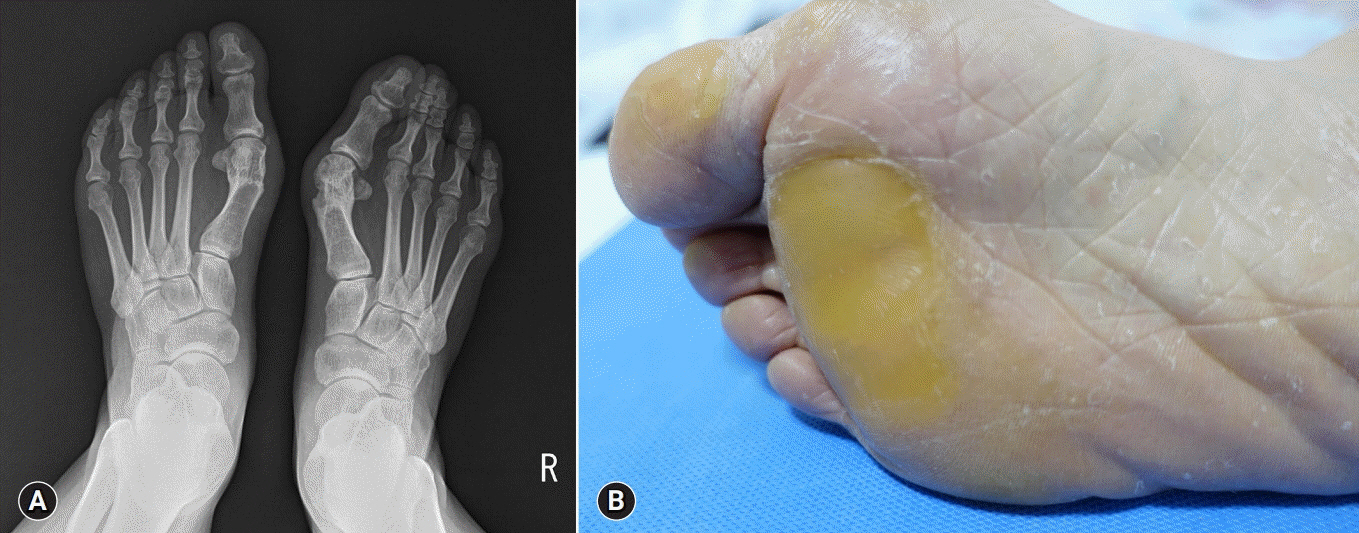
Fig. 1. (A) Metatarsal shortening of first metatarsal on right foot due to hallux valgus recurrence after hallux valgus surgery. (B) Callosity under second and third metatarsal head due to altered metatarsal position after hallux valgus surgery.

Fig. 2. Orthosis for treatment of hallux valgus. (A) Toe spreader, (B) Valgus splint, and (C) Bunion shield.

Fig. 3. Hammer toe with a flexion deformity at the proximal interphalangeal joint of the toe accompanied by a slight metatarsophalangeal joint extension deformity.
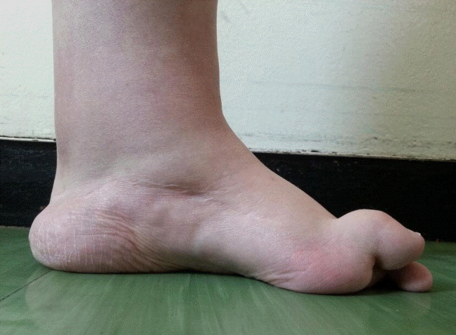
Fig. 4. Claw toe with a hyperextension deformity at the metatarsophalangeal joint and secondarily having flexion deformity in the proximal interphalangeal and distal interphalangeal joints.
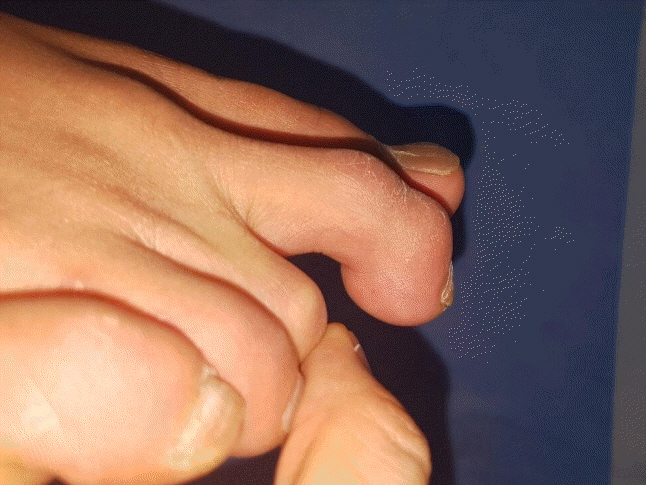
Fig. 5. Mallet toe with a flexion deformity at the distal interphalangeal joint.
Reference
-
References
1. Chang MC. Reduced foot pain after spasticity control with alcohol block in a patient with chronic hemiparetic stroke: a case report. J Phys Ther Sci. 2017; 29:767–70.
Article2. Chang MC. Metatarsalgia in a patient with chronic hemiparetic stroke managed with alcohol block of the tibial nerve: a case report. Neurol Asia. 2017; 22:267–70.3. Branthwaite H, Chockalingam N, Greenhalgh A. The effect of shoe toe box shape and volume on forefoot interdigital and plantar pressures in healthy females. J Foot Ankle Res. 2013; 6:28.
Article4. Martorell JM. Hallux disorder and metatarsal alignment. Clin Orthop Relat Res. 1981; (157):14–20.
Article5. DiPreta JA. Metatarsalgia, lesser toe deformities, and associated disorders of the forefoot. Med Clin North Am. 2014; 98:233–51.
Article6. Espinosa N, Brodsky JW, Maceira E. Metatarsalgia. J Am Acad Orthop Surg. 2010; 18:474–85.
Article7. Bardelli M, Turelli L, Scoccianti G. Definition and classification of metatarsalgia. Foot and Ankle Surg. 2003; 9:79–85.
Article8. Charen DA, Markowitz JS, Cheung ZB, Matijakovich DJ, Chan JJ, Vulcano E. Overview of metatarsalgia. Orthopedics. 2019; 42:e138–43.
Article9. Maestro M, Besse JL, Ragusa M, Berthonnaud E. Forefoot morphotype study and planning method for forefoot osteotomy. Foot Ankle Clin. 2003; 8:695–710.
Article10. Slullitel G, López V, Calvi JP, Seletti M, Bartolucci C, Pinton G. Effect of first ray insufficiency and metatarsal index on metatarsalgia in hallux valgus. Foot Ankle Int. 2016; 37:300–6.
Article11. Espinosa N, Maceira E, Myerson MS. Current concept review: metatarsalgia. Foot Ankle Int. 2008; 29:871–9.
Article12. Gauthier G, Elbaz R. Freiberg's infraction: a subchondral bone fatigue fracture. A new surgical treatment. Clin Orthop Relat Res. 1979; (142):93–5.13. Talusan PG, Diaz-Collado PJ, Reach JS Jr. Freiberg's infraction: diagnosis and treatment. Foot Ankle Spec. 2014; 7:52–6.14. Acevedo JI. Fixation of metatarsal osteotomies in the treatment of hallux valgus. Foot Ankle Clin. 2000; 5:451–68.15. Hofstaetter SG, Hofstaetter JG, Petroutsas JA, Gruber F, Ritschl P, Trnka HJ. The Weil osteotomy: a seven-year follow-up. J Bone Joint Surg Br. 2005; 87:1507–11.16. Reddy VB. Metatarsal osteotomies: complications. Foot Ankle Clin. 2018; 23:47–55.17. Chang AH, Abu-Faraj ZU, Harris GF, Nery J, Shereff MJ. Multistep measurement of plantar pressure alterations using metatarsal pads. Foot Ankle Int. 1994; 15:654–60.
Article18. Hsi WL, Kang JH, Lee XX. Optimum position of metatarsal pad in metatarsalgia for pressure relief. Am J Phys Med Rehabil. 2005; 84:514–20.
Article19. Männikkö K, Sahlman J. The effect of metatarsal padding on pain and functional ability in metatarsalgia. Scand J Surg. 2017; 106:332–7.
Article20. Robinson AH, Limbers JP. Modern concepts in the treatment of hallux valgus. J Bone Joint Surg Br. 2005; 87:1038–45.
Article21. Nix S, Smith M, Vicenzino B. Prevalence of hallux valgus in the general population: a systematic review and meta-analysis. J Foot Ankle Res. 2010; 3:21.
Article22. Hecht PJ, Lin TJ. Hallux valgus. Med Clin North Am. 2014; 98:227–32.
Article23. McBride ED. A conservative operation for bunions. J Bone Joint Surg Am. 1928; 10:735–9.
Article24. Perera AM, Mason L, Stephens MM. The pathogenesis of hallux valgus. J Bone Joint Surg Am. 2011; 93:1650–61.
Article25. Nery C, Coughlin MJ, Baumfeld D, Ballerini FJ, Kobata S. Hallux valgus in males--part 1: demographics, etiology, and comparative radiology. Foot Ankle Int. 2013; 34:629–35.26. Wilkerson RD, Mason MA. Differences in men's and women's mean ankle ligamentous laxity. Iowa Orthop J. 2000; 20:46–8.27. Tehraninasr A, Saeedi H, Forogh B, Bahramizadeh M, Keyhani MR. Effects of insole with toe-separator and night splint on patients with painful hallux valgus: a comparative study. Prosthet Orthot Int. 2008; 32:79–83.
Article28. Cha YH, Kim SJ, Lee KH, Kwon JY, Kim DH, Seo A, et al. Designing personalized toe spreaders for hallux valgus with three-dimensional scanning and printing. J Biomed Eng Biosci. 2018; 5:1–6.
Article29. Camasta CA. Hallux limitus and hallux rigidus. Clinical examination, radiographic findings, and natural history. Clin Podiatr Med Surg. 1996; 13:423–48.30. Coughlin MJ, Shurnas PS. Hallux rigidus: demographics, etiology, and radiographic assessment. Foot Ankle Int. 2003; 24:731–43.
Article31. Shereff MJ, Baumhauer JF. Hallux rigidus and osteoarthrosis of the first metatarsophalangeal joint. J Bone Joint Surg Am. 1998; 80:898–908.
Article32. Beeson P, Phillips C, Corr S, Ribbans W. Classification systems for hallux rigidus: a review of the literature. Foot Ankle Int. 2008; 29:407–14.
Article33. Bingold AC, Collins DH. Hallux rigidus. J Bone Joint Surg Br. 1950; 32:214–22.
Article34. Zammit GV, Menz HB, Munteanu SE. Structural factors associated with hallux limitus/rigidus: a systematic review of case control studies. J Orthop Sports Phys Ther. 2009; 39:733–42.
Article35. Bartlett DH. Arthroscopic management of osteochondritis dissecans of the first metatarsal head. Arthroscopy. 1988; 4:51–4.
Article36. Bonney G, macnab I. Hallux valgus and hallux rigidus; a critical survey of operative results. J Bone Joint Surg Br. 1952; 34:366–85.37. Shrader JA, Siegel KL. Nonoperative management of functional hallux limitus in a patient with rheumatoid arthritis. Phys Ther. 2003; 83:831–43.
Article38. Schuh R, Trnka HJ. First metatarsophalangeal arthrodesis for severe bone loss. Foot Ankle Clin. 2011; 16:13–20.
Article39. Becerro de Bengoa Vallejo R, Sanchez Gómez R, Losa Iglesias ME. Clinical improvement in functional hallux limitus using a cut-out orthosis. Prosthet Orthot Int. 2016; 40:215–23.
Article40. Rambarran KK, Lemaire E, Robertson GD. Effectiveness of the Kinetic Wedge foot orthosis modification to reduce relative plantar pressure. 25th Conference of the American Society of Biomechanics [Internet]. 2003. [cited 2019 March 28]. http://www.asbweb.org/conferences/2003/pdfs/163.pdf.41. Menz HB, Levinger P, Tan JM, Auhl M, Roddy E, Munteanu SE. Rocker-sole footwear versus prefabricated foot orthoses for the treatment of pain associated with first metatarsophalangeal joint osteoarthritis: study protocol for a randomised trial. BMC Musculoskelet Disord. 2014; 15:86.
Article42. Smith DG, Barnes BC, Sands AK, Boyko EJ, Ahroni JH. Prevalence of radiographic foot abnormalities in patients with diabetes. Foot Ankle Int. 1997; 18:342–6.
Article43. Malhotra K, Davda K, Singh D. The pathology and management of lesser toe deformities. EFORT Open Rev. 2017; 1:409–19.
Article44. Stainsby GD. Pathological anatomy and dynamic effect of the displaced plantar plate and the importance of the integrity of the plantar plate-deep transverse metatarsal ligament tie-bar. Ann R Coll Surg Engl. 1997; 79:58–68.45. Shirzad K, Kiesau CD, DeOrio JK, Parekh SG. Lesser toe deformities. J Am Acad Orthop Surg. 2011; 19:505–14.
Article46. Coughlin MJ, Dorris J, Polk E. Operative repair of the fixed hammertoe deformity. Foot Ankle Int. 2000; 21:94–104.
Article47. Mizel MS, Michelson JD. Nonsurgical treatment of monarticular nontraumatic synovitis of the second metatarsophalangeal joint. Foot Ankle Int. 1997; 18:424–6.
Article48. Di Caprio F, Meringolo R, Shehab Eddine M, Ponziani L. Morton's interdigital neuroma of the foot: a literature review. Foot Ankle Surg. 2018; 24:92–8.49. Bradley N, Miller WA, Evans JP. Plantar neuroma: analysis of results following surgical excision in 145 patients. South Med J. 1976; 69:853–4.50. Kasparek M, Schneider W. Surgical treatment of Morton's neuroma: clinical results after open excision. Int Orthop. 2013; 37:1857–61.
Article51. Sofka CM, Adler RS, Ciavarra GA, Pavlov H. Ultrasound-guided interdigital neuroma injections: short-term clinical outcomes after a single percutaneous injection--preliminary results. HSS J. 2007; 3:44–9.
Article52. Climent JM, Mondéjar-Gómez F, Rodríguez-Ruiz C, Díaz-Llopis I, Gómez-Gallego D, Martín-Medina P. Treatment of Morton neuroma with botulinum toxin A: a pilot study. Clin Drug Investig. 2013; 33:497–503.
Article53. Hinz A. Nerve disorders. In : DiGiovanni CW, Greisberg J, editors. Foot and ankle: core knowledge in orthopaedics. Philadelphia: Elsevier Mosby;2007. p. 171–6.
