Large Forefoot Schwannoma: A Case Report
- Affiliations
-
- 1Department of Orthopaedic Surgery, National Medical Center, Seoul, Korea. mdjsh@nate.com
- KMID: 2181644
- DOI: http://doi.org/10.14193/jkfas.2014.18.4.212
Abstract
- A schwannoma is a benign neurogenic tumor derived from Schwann cells. A rare case of a large painful schwannoma in the foot with metatarsal deformity was presented. Due to suspicion of malignancy, amputation had been recommended previously. We report on a rare case of a large forefoot schwannoma causing pain and paresthesia of the forefoot.
Keyword
Figure
-
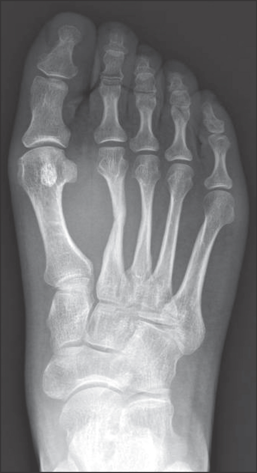
Figure 1. Anteroposterior radiograph shows lateral angulation and sclerosis of second, third metatarsal shaft.
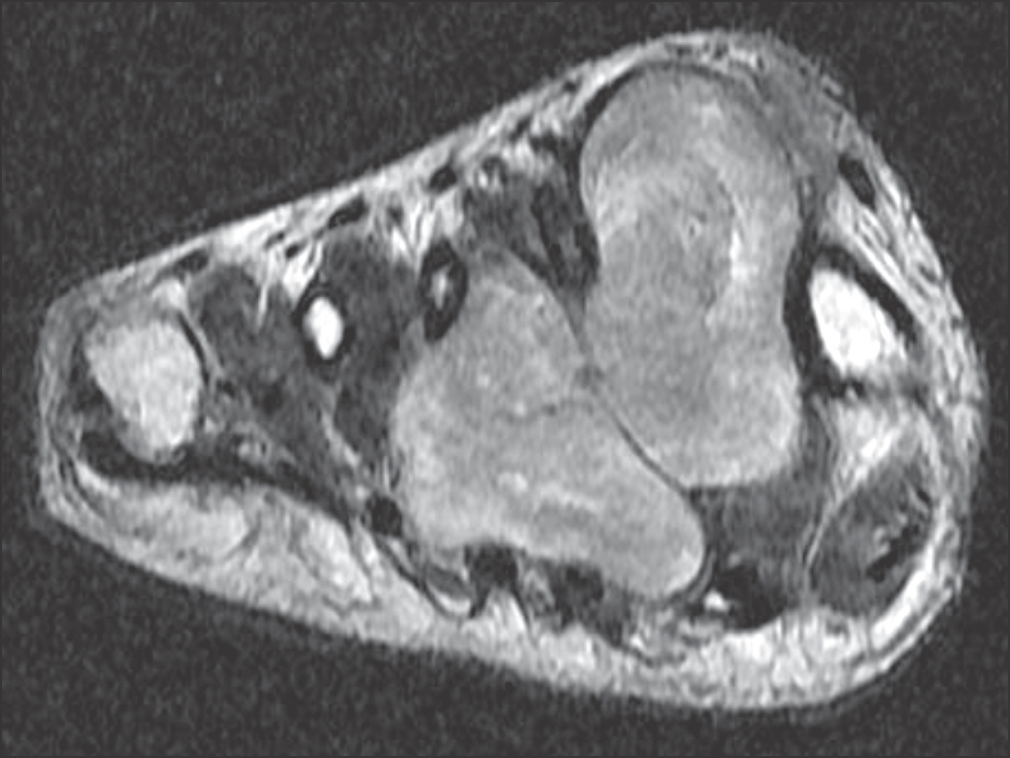
Figure 2. The axial T2-weighted magnetic resonance image shows a heterogenous cystic mass with low to hyperdense signal intensity, with dorsal and plantar extension.
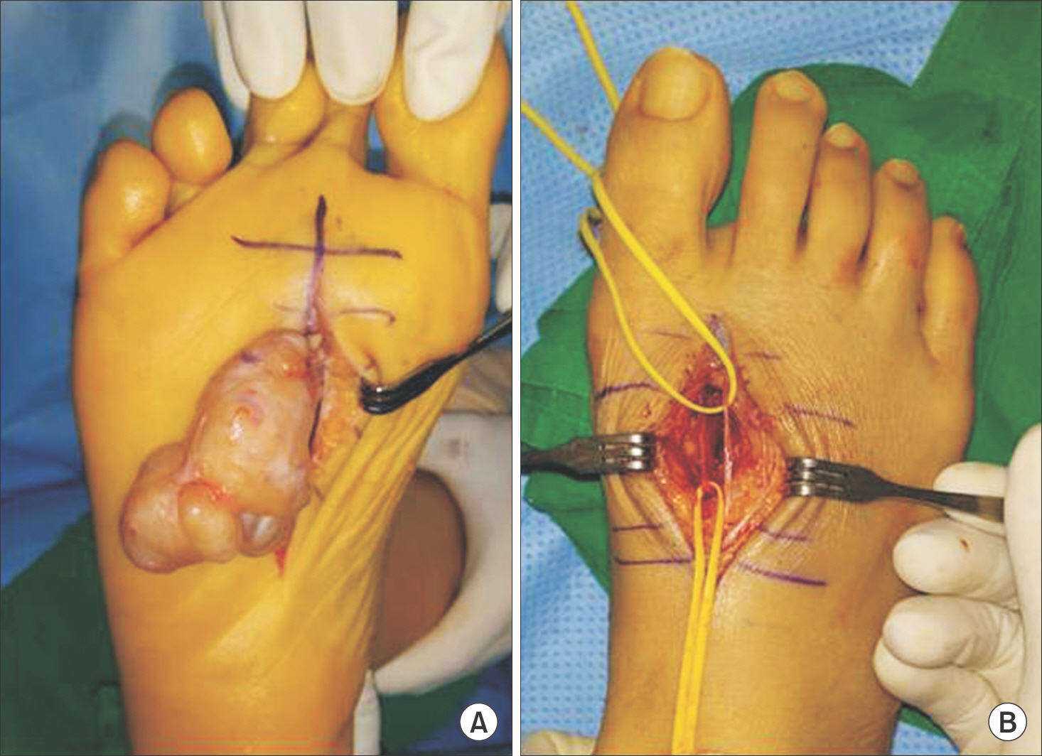
Figure 3. (A) A excised mass was removed through plantar incision. (B) Clinical photograph after mass excision shows both a deep peroneal nerve and a common proper digital nerve derived from medial plantar nerve.

Figure 4. (A) Gross photograph shows a well circumscribed and encapsulated mass. (B) The cross section of mass appears diffusely myxoid and grayish yellow cut surfaces without hemorrhage or necrosis.
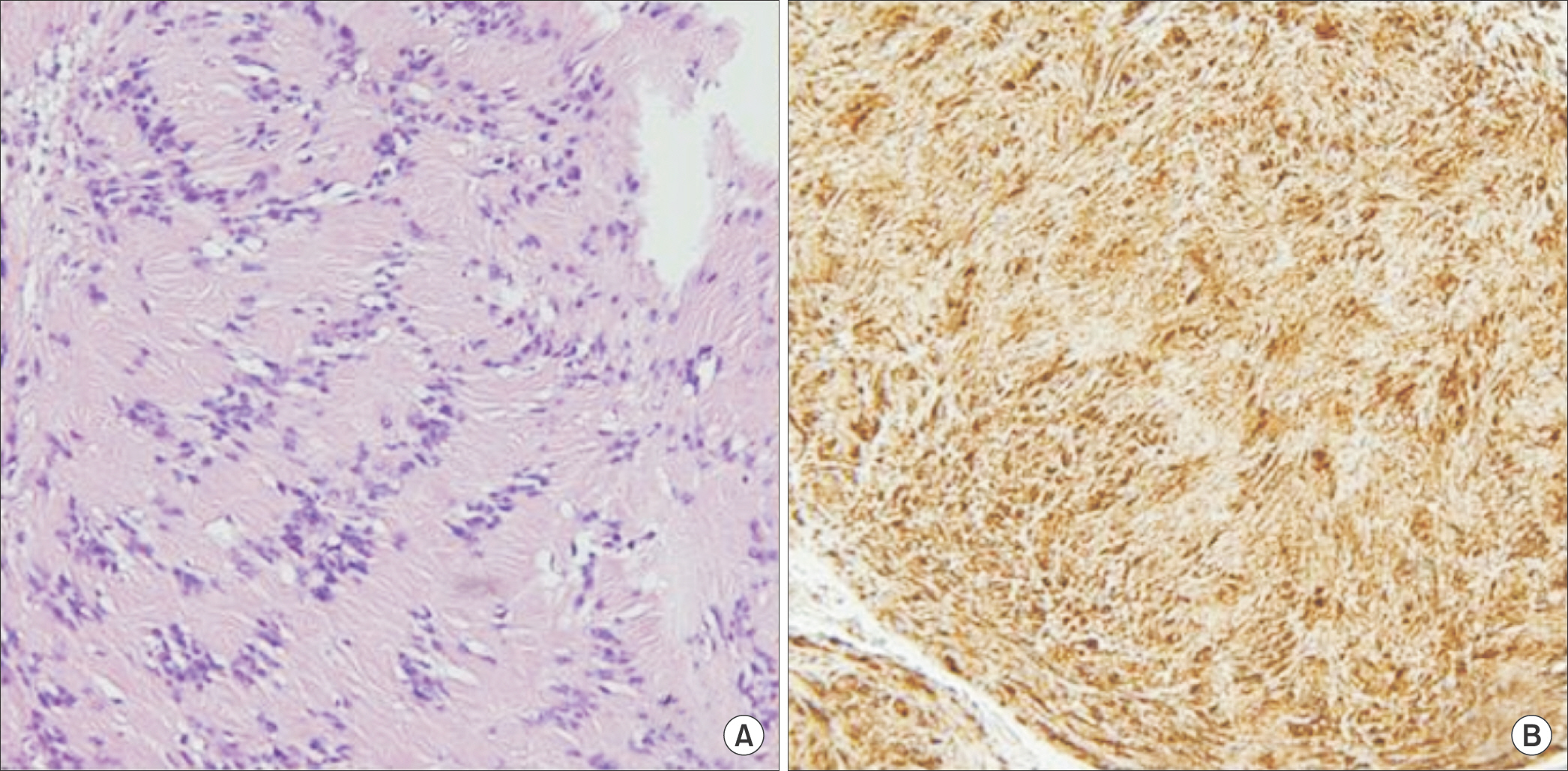
Figure 5. (A) Histological features show a characteristic biphasic pattern of Antoni A and Antoni B of neurogenic spindle cell proliferation (H&E stain, ×100). (B) Immunostaining for the S-100 protein was strong and diffuse confirming the diagnosis of schwannoma (×100).
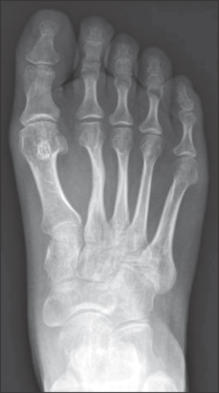
Figure 6. Anteroposterior radiograph shows a remodeling of second, third metatarsal shaft six month later.
Reference
-
1.Enzinger FM., Weiss SW. Benign tumors of peripheral nerves. Enzinger FM, Weiss SW, editors. editors.Enzinger and Weiss’s soft tissue tumors. 5th ed.St Louis: Mosby;2008. p. 853–69.2.Weiss SW., Langloss JM., Enzinger FM. Value of S-100 protein in the diagnosis of soft tissue tumors with particular reference to benign and malignant Schwann cell tumors. Lab Invest. 1983. 49:299–308.3.Mangrulkar VH., Brunetti VA., Gould ES., Howell N. Unusually large pedal schwannoma. J Foot Ankle Surg. 2007. 46:398–402.
Article4.Pyun YS., Kim SR., Joh YR. Surgical treatment of the neurilemoma in extremities. J Korean Bone Joint Tumor Soc. 1998. 4:88–93.5.Das Gupta TK., Brasfield RD., Strong EW., Hajdu SI. Benign solitary Schwannomas (neurilemomas). Cancer. 1969. 24:355–66.6.Odom RD., Overbeek TD., Murdoch DP., Hosch JC. Neurilemoma of the medial plantar nerve: a case report and literature review. J Foot Ankle Surg. 2001. 40:105–9.
Article7.Rettenbacher T., Sögner P., Springer P., Fiegl M., Hussl H., zur Ned-den D. Schwannoma of the brachial plexus: cross-sectional imaging diagnosis using CT, sonography, and MR imaging. Eur Radiol. 2003. 13:1872–5.
Article8.Lee SH., Jung HG., Lee HK. Neurilemoma of trunk and extremities. J Korean Orthop Assoc. 1996. 31:556–63.
Article9.Flores Santos F., Pinheiro M., Felicíssimo P. Large foot schwannoma with bone invasion: a case report. Foot Ankle Surg. 2014. 20:e23–6.
- Full Text Links
-
- Actions
-
Cited
- CITED
-
- Close
- Share
-
- Similar articles
-
- Pigmented Villonodular Synovitis in Forefoot (A Case Report)
- Hypervascular Vestibular Schwannoma: A Case Report
- A Case of Pelvic Retroperitoneal Schwannoma : Preoperatively Suspected of Malignant Adnexal Tumor
- Melanotic Schwannoma in Cervical Spine: A Case Report
- A Case Report of Large Schwannoma Arising in Sciatic Nerve
