Hypervascular Vestibular Schwannoma: A Case Report
- Affiliations
-
- 1Department of Radiology, Eulji University Hospital, Daejeon, Korea. midosyu@eulji.ac.kr
- KMID: 2208788
- DOI: http://doi.org/10.3348/jksr.2014.71.5.197
Abstract
- Most vestibular schwannoma is hypovascular with well known poor tumor staining in cerebral angiography. However, hypervascular vestibular schwannoma might be observed as a rare subtype with increased risk of bleeding during surgery. Multimodal imaging features which represent hypervascularity of the tumor can be observed in hypervascular vestibular schwannoma. Here we report a case of hypervascular vestibular schwannoma with brief literature review.
MeSH Terms
Figure
-
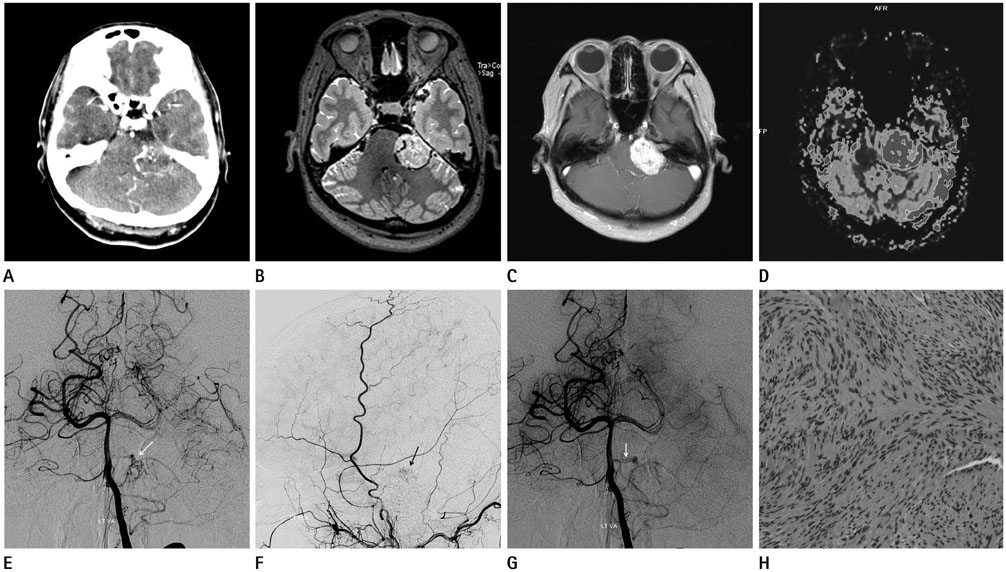
Fig. 1 A 52-year-old female with hypervascular vestibular schwannoma. A. Early arterial enhanced CT reveals prominent enhancing vessels in and around the tumor. B. T2-weighted image shows intratumoral and peritumoral signal void structures. C. Enhanced T1-weighted image shows inhomogeneous strong enhancement with focal internal auditory canal tumor portion. D. Perfusion MRI shows markedly increased relative cerebral blood volume of the left cerebellopontine angle mass. E-G. Cerebral angiography reveals early tumor staining, supplied by the left anterior inferior cerebellar artery and by the left middle meningeal artery (arrows) (E, F). And engorged draining vein with early venous filling is also seen (arrow) (G). H. Hematoxylin and eosin staining (× 200) shows cellular pattern with strongly stained nuclear palisading with hyaline vessel walls.
Reference
-
1. Nikolopoulos TP, Fortnum H, O'Donoghue G, Baguley D. Acoustic neuroma growth: a systematic review of the evidence. Otol Neurotol. 2010; 31:478–485.2. Beaman FD, Kransdorf MJ, Menke DM. Schwannoma: radiologic-pathologic correlation. Radiographics. 2004; 24:1477–1481.3. Yamakami I, Kobayashi E, Iwadate Y, Saeki N, Yamaura A. Hypervascular vestibular schwannomas. Surg Neurol. 2002; 57:105–112.4. Han L, Shu K, Guo D, Lei T, Li L. Hypervascular acoustic tumor. Chinese-German. J Clin Oncol. 2006; 5:366–368.5. LeMay DR, Sun JK, Fishback D, Locke GE, Giannotta SL. Hypervascular acoustic neuroma. Neurol Res. 1998; 20:748–750.6. Arienta C, Caroli M, Crotti FM. Subarachnoid haemorrhage due to acoustic neurinoma Case report and review of the literature. Neurochirurgia (Stuttg). 1988; 31:162–165.7. Baba M, Iseki H, Kumagai Y, Sugiyama H, Nawada H. [Acoustic neurinoma with massive hemorrhage within the tumor tissue--a case report (author's transl)]. No Shinkei Geka. 1980; 8:193–197.8. Vellin JF, Bozorg Grayeli A, Kalamarides M, Fond C, Bouccara D, Sterkers O. Intratumoral and brainstem hemorrhage in a patient with vestibular schwannoma and oral anticoagulant therapy. Otol Neurotol. 2006; 27:209–212.
- Full Text Links
-
- Actions
-
Cited
- CITED
-
- Close
- Share
-
- Similar articles
-
- Diagnosis and Management of Vestibular Schwannoma: Focus on Dizziness
- Normal pressure hydrocephalus after gamma knife radiosurgery in a patient with vestibular schwannoma
- A Case of Intralabyrinthine Schwannoma and Literature Review of the Cases Reported Previously in Korea
- A Case of Marked Hearing Improvement after Surgical Removal of Vestibular Schwannoma with Profound Hearing Loss
- A Case of inferior vestibular schwannoma which was lately diagnosed due to normal hearing level
