Brain Areas Subserving Torrance Tests of Creative Thinking: An Functional Magnetic Resonance Imaging Study
- Affiliations
-
- 1Department of Neurology, Dongguk University Ilsan Hospital, Goyang, Korea. neukim@duih.org
- 2Department of Education, Dongguk University, Seoul, Korea.
- KMID: 2442826
- DOI: http://doi.org/10.12779/dnd.2017.16.2.48
Abstract
- BACKGROUND AND PURPOSE
Torrance Tests of Creative Thinking (TTCT) is a well-known and commonly used measure of creativity. However, the TTCT-induced creative hemodynamic brain activity is rarely revealed. The purpose of this study is to elucidate the neural correlates of creative thinking in the setting of a modified version of the figural TTCT adapted for an functional magnetic resonance imaging (fMRI) experiment.
METHODS
We designed a blocked fMRI experiment. Twenty-five participants (11 males, 14 females, mean age 19.9±1.8) were asked to complete the partially presented line drawing of the figural TTCT (creative drawing imagery; creative). As a control condition, subjects were asked to keep tracking the line on the screen (line tracking; control).
RESULTS
Compared to the control condition, creative condition revealed greater activation in the distributed and bilateral brain regions including the left anterior cingulate, bilateral frontal, parietal, temporal and occipital regions as shown in the previous creativity studies.
CONCLUSIONS
The present revealed the neural basis underlying the figural TTCT using fMRI, providing an evidence of brain areas encompassing the figural TTCT. Considering the significance of a creativity test for dementia patients, the neural correlates of TTCT elucidated by this study may be valuable to evaluate the brain function of patients in the clinical field.
MeSH Terms
Figure
-
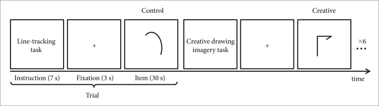
Fig. 1 fMRI experimental paradigm for creative thinking using the figural TTCT. A modified figural TTCT was used to examine brain regions related to visual creativity using fMRI. fMRI experiments consisted of the two conditions: creative drawing imagery (creative) as an active condition and line-tracking (control) as a control condition. For each condition there were six trials, where an instruction was presented for 7 s, a fixation for 3 s, and an item for 30 s subsequently. The items for creative condition were from the figural TTCT form B while that for control condition were novel items devised for this study. fMRI: functional magnetic resonance imaging, TTCT: Torrance Tests of Creative Thinking.
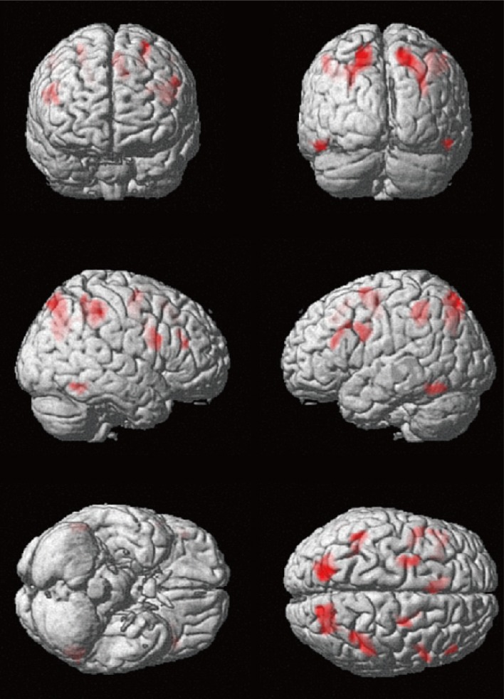
Fig. 2 Brain regions revealing greater activation during creative drawing imagery compared to line tracking condition. Group analysis revealed brain activation for creative drawing imagery condition than line tracking condition (creative>control) (p<0.001, uncorrected for multiple comparisons; cluster size≥50). The threshold activations were revealed on 3D brain images by projecting results on to the surface of the brain. 3D: 3-dimensional.
Reference
-
1. Runco MA, Jaeger GJ. The standard definition of creativity. Crea Res J. 2012; 24:92–96.
Article2. Fink A, Benedek M, Grabner RH, Staudt B, Neubauer AC. Creativity meets neuroscience: experimental tasks for the neuroscientific study of creative thinking. Methods. 2007; 42:68–76. PMID: 17434417.
Article3. Arden R, Chavez RS, Grazioplene R, Jung RE. Neuroimaging creativity: a psychometric view. Behav Brain Res. 2010; 214:143–156. PMID: 20488210.
Article4. Torrance EP.Torrance Tests of Creative Thinking: norms-technical manual: figural (streamlined) forms A & B. Bensenville: Scholastic Testing Service;1998.5. Piffer D. Can creativity be measured? An attempt to clarify the notion of creativity and general directions for future research. Think Skills Creat. 2012; 7:258–264.
Article6. Kim KH. Can we trust creativity tests? A review of the Torrance Tests of Creative Thinking (TTCT). Creat Res J. 2006; 18:3–14.
Article7. Palmiero M, Di Giacomo D, Passafiume D. Creativity and dementia: a review. Cogn Process. 2012; 13:193–209. PMID: 22438178.
Article8. Dietrich A, Kanso R. A review of EEG, ERP, and neuroimaging studies of creativity and insight. Psychol Bull. 2010; 136:822–848. PMID: 20804237.
Article9. Cramond B. The Torrance Tests of Creative Thinking: from design through establishment of predictive validity. In : Subotnik RF, Arnold KD, editors. Beyond terman: contemporary longitudinal studies of giftedness and talent. Norwood: Ablex;1994. p. 229–254.10. Wiggins GA, Bhattacharya J. Mind the gap: an attempt to bridge computational and neuroscientific approaches to study creativity. Front Hum Neurosci. 2014; 8:540. PMID: 25104930.
Article11. Runco MA, Yoruk S. The neuroscience of divergent thinking. Act Nerv Super. 2014; 56:1–16.
Article12. Oldfield RC. The assessment and analysis of handedness: the Edinburgh inventory. Neuropsychologia. 1971; 9:97–113. PMID: 5146491.
Article13. Lancaster JL, Woldorff MG, Parsons LM, Liotti M, Freitas CS, Rainey L, et al. Automated Talairach atlas labels for functional brain mapping. Hum Brain Mapp. 2000; 10:120–131. PMID: 10912591.
Article14. Lancaster JL, Rainey LH, Summerlin JL, Freitas CS, Fox PT, Evans AC, et al. Automated labeling of the human brain: a preliminary report on the development and evaluation of a forward-transform method. Hum Brain Mapp. 1997; 5:238–242. PMID: 20408222.
Article15. Lancaster JL, Tordesillas-Gutiérrez D, Martinez M, Salinas F, Evans A, Zilles K, et al. Bias between MNI and Talairach coordinates analyzed using the ICBM-152 brain template. Hum Brain Mapp. 2007; 28:1194–1205. PMID: 17266101.
Article16. Mechelli A, Price CJ, Friston KJ, Ishai A. Where bottom-up meets top-down: neuronal interactions during perception and imagery. Cereb Cortex. 2004; 14:1256–1265. PMID: 15192010.
Article17. Ganis G, Thompson WL, Kosslyn SM. Brain areas underlying visual mental imagery and visual perception: an fMRI study. Brain Res Cogn Brain Res. 2004; 20:226–241. PMID: 15183394.
Article18. De Pisapia N, Bacci F, Parrott D, Melcher D. Brain networks for visual creativity: a functional connectivity study of planning a visual artwork. Sci Rep. 2016; 6:39185. PMID: 27991592.
Article19. Davis KD, Hutchison WD, Lozano AM, Tasker RR, Dostrovsky JO. Human anterior cingulate cortex neurons modulated by attention-demanding tasks. J Neurophysiol. 2000; 83:3575–3577. PMID: 10848573.
Article20. Huang P, Qiu L, Shen L, Zhang Y, Song Z, Qi Z, et al. Evidence for a left-over-right inhibitory mechanism during figural creative thinking in healthy nonartists. Hum Brain Mapp. 2013; 34:2724–2732. PMID: 22522783.
Article21. Aziz-Zadeh L, Liew SL, Dandekar F. Exploring the neural correlates of visual creativity. Soc Cogn Affect Neurosci. 2013; 8:475–480. PMID: 22349801.
Article22. Jahanshahi M, Dirnberger G. The left dorsolateral prefrontal cortex and random generation of responses: studies with transcranial magnetic stimulation. Neuropsychologia. 1999; 37:181–190. PMID: 10080375.
Article23. Pollmann S, von Cramon DY. Object working memory and visuospatial processing: functional neuroanatomy analyzed by event-related fMRI. Exp Brain Res. 2000; 133:12–22. PMID: 10933206.
Article24. Hampshire A, Owen AM. Fractionating attentional control using event-related fMRI. Cereb Cortex. 2006; 16:1679–1689. PMID: 16436686.
Article25. Boroojerdi B, Phipps M, Kopylev L, Wharton CM, Cohen LG, Grafman J. Enhancing analogic reasoning with rTMS over the left prefrontal cortex. Neurology. 2001; 56:526–528. PMID: 11222799.
Article26. Hanakawa T, Honda M, Sawamoto N, Okada T, Yonekura Y, Fukuyama H, et al. The role of rostral Brodmann area 6 in mental-operation tasks: an integrative neuroimaging approach. Cereb Cortex. 2002; 12:1157–1170. PMID: 12379604.
Article27. Ardila A, Bernal B, Rosselli M. Language and visual perception associations: meta-analytic connectivity modeling of Brodmann area 37. Behav Neurol. 2015; 2015:565871. PMID: 25648869.
Article28. Luria AR. Basic Problems of Neurolinguistics. Hague: Walter de Gruyter;1976.29. Berthier ML. Transcortical Aphasias. Hove: Psychology Press;1999.
- Full Text Links
-
- Actions
-
Cited
- CITED
-
- Close
- Share
-
- Similar articles
-
- Functional Magnetic Resonance Imaging of the Brain: Principle and Practical Application
- Application of Functional MRI in the Field of Rehabilitation Medicine
- Neuro-Scientific Studies of Creativity
- Advanced Magnetic Resonance Imaging for Pediatric Brain Tumors: Current Imaging Techniques and Interpretation Algorithms
- Advanced Imaging of Traumatic Brain Injury
