A Case of Hepatic Inflammatory Pseudotumor Occurred in a Patient with Lupus Nephritis
- Affiliations
-
- 1Department of Rheumatology, Hanyang University Hospital for Rheumatic Diseases, Seoul, Korea. sungyk@hanyang.ac.kr
- 2Department of Internal Medicine, Hanyang University College of Medicine, Seoul, Korea.
- KMID: 2442025
- DOI: http://doi.org/10.4078/jrd.2019.26.2.137
Abstract
- Systemic lupus erythematosus (SLE) is a systemic autoimmune disease affecting various organs. Among its manifestations, inflammatory pseudotumor (IPT) is an extremely rare disease about which no case has been reported of it occurring in the liver. We present a case of a SLE patient with hepatic IPT (hIPT) successfully treated with immunosuppressants. A 16-year-old male with elevated liver enzymes visited our clinic and was diagnosed as SLE. Although no lesion was observed in the initial abdomen ultrasonography, the abdominal CT on hospital day 7 revealed a new hepatic mass resembling an abscess. Despite 5 weeks of antibiotics treatment, the hepatic mass remained, and was re-diagnosed as hIPT secondary to SLE with an abdominal MRI. After high dose prednisolone and mycophenolate mofetil treatment, lupus activity subsided and hIPT disappeared in the follow-up CT. This case suggests that hIPT should be considered as a differential diagnosis among hepatic mass in SLE patients.
MeSH Terms
-
Abdomen
Abscess
Adolescent
Anti-Bacterial Agents
Autoimmune Diseases
Diagnosis, Differential
Follow-Up Studies
Granuloma
Granuloma, Plasma Cell*
Humans
Immunosuppressive Agents
Liver
Liver Neoplasms
Lupus Erythematosus, Systemic
Lupus Nephritis*
Magnetic Resonance Imaging
Male
Plasma Cells
Prednisolone
Rare Diseases
Tomography, X-Ray Computed
Ultrasonography
Anti-Bacterial Agents
Immunosuppressive Agents
Prednisolone
Figure
-
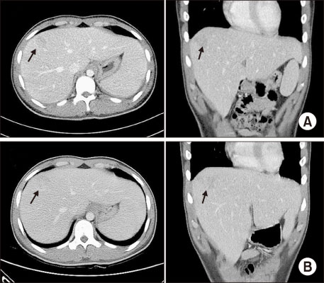
Figure 1 (A) Initial contrast enhanced abdominopelvic computed tomography (CT) on hospital day (HD) 7 with transverse (left) and coronal (right) view showing rim-enhancing hypo-attenuated lesion at subcapsular area, largest lesion (arrows) measured 3.4×2.5 cm on segment 8. (B) Follow-up CT on HD 18 with transverse (left) and coronal (right) view revealing unchanged hepatic lesion.

Figure 2 Follow-up transverse view of liver magnetic resonance imaging images on 5 weeks after antibiotics treatment, with (A) Axial T2-weighted fast spin echo, (B) diffusion weighted image on b=800 s/mm2, (C) apparent diffusion coefficient map, (D~F) portal-venous, equilibrium, and hepatobiliary phase. The largest lesion measured 2.6×2.0 cm on segment 8.

Figure 3 Follow-up of abdominopelvic contrast-enhanced computed tomography. The previous lesion on liver segment 8 was completely resolved in axial (A) and coronal view (B) of portal phase.

Figure 4 Clinical course of the laboratory findings (A) and the medications (B). AST: aspartate aminotransferase, ALT: alanine aminotransferase, ESR: erythrocyte sedimentation rate, CRP: C-reactive protein, C3: complement 3, C4: complement 4, PDS: prednisolone, MMF: Mycophenolate mofetil.
Reference
-
1. Patnana M, Sevrukov AB, Elsayes KM, Viswanathan C, Lubner M, Menias CO. Inflammatory pseudotumor: the great mimicker. AJR Am J Roentgenol. 2012; 198:W217–W227.
Article2. Ntinas A, Kardassis D, Miliaras D, Tsinoglou K, Dimitriades A, Vrochides D. Inflammatory pseudotumor of the liver: a case report and review of the literature. J Med Case Rep. 2011; 5:196.
Article3. Díaz-torné C, Narváez J, De Lama E, Diez-García M, Narváez JA, Bernad B, et al. Inflammatory pseudotumor of the liver associated with rheumatoid arthritis. Arthritis Rheum. 2007; 57:1102–1106.
Article4. Younis N, Khaleeli AA, Soran H, Monteith PG. Inflammatory pseudotumour of the liver associated with diabetes mellitus. Int J Clin Pract. 2001; 55:717–719.5. Bessone F, Poles N, Roma MG. Challenge of liver disease in systemic lupus erythematosus: clues for diagnosis and hints for pathogenesis. World J Hepatol. 2014; 6:394–409.
Article6. Khatri A, Agrawal A, Sikachi RR, Mehta D, Sahni S, Meena N. Inflammatory myofibroblastic tumor of the lung. Adv Respir Med. 2018; 86:27–35.
Article7. Oh JS, Kwon GY, So MW, Choi SH, Kim YG, Nah SS, et al. A case of plasma cell granuloma of skull in a patient with systemic lupus erythematosus. J Korean Rheum Assoc. 2006; 13:311–315.8. Piga M, Vacca A, Porru G, Cauli A, Mathieu A. Liver involvement in systemic lupus erythematosus: incidence, clinical course and outcome of lupus hepatitis. Clin Exp Rheumatol. 2010; 28:504–510.9. Chowdhary VR, Crowson CS, Poterucha JJ, Moder KG. Liver involvement in systemic lupus erythematosus: case review of 40 patients. J Rheumatol. 2008; 35:2159–2164.
Article10. Park JY, Choi MS, Lim YS, Park JW, Kim SU, Min YW, et al. Clinical features, image findings, and prognosis of inflammatory pseudotumor of the liver: a multicenter experience of 45 cases. Gut Liver. 2014; 8:58–63.
Article11. Wangkaew S, Lertprasertsuk N, Chotirosniramit A, Louthrenoo W. Hepatic vasculitis presenting with multiple sterile liver abscesses in a patient with systemic lupus erythematosus. Int J Rheum Dis. 2007; 10:64–68.
Article12. Syed MA, Kim TK, Jang HJ. Portal and hepatic vein thrombosis in liver abscess: CT findings. Eur J Radiol. 2007; 61:513–519.
Article13. Thietart S, Mekinian A, Delorme S, Lequoy M, Gobert D, Arrivé L, et al. Vasculitis of the hepatic artery: a case of a single-organ vasculitis. Rev Med Interne. 2017; 38:847–849.14. Seo N, Kim DY, Choi JY. Cross-sectional imaging of intrahepatic cholangiocarcinoma: development, growth, spread, and prognosis. AJR Am J Roentgenol. 2017; 209:W64–W75.
Article15. Yan FH, Zhou KR, Jiang YP, Shi WB. Inflammatory pseudotumor of the liver: 13 cases of MRI findings. World J Gastroenterol. 2001; 7:422–424.
Article
- Full Text Links
-
- Actions
-
Cited
- CITED
-
- Close
- Share
-
- Similar articles
-
- A case of Lupus Nephritis
- A Case of Pseudotumor Cerebri Associated with Systemic Lupus Erythematosus
- A Case of Lupus Nephritis in a Patient with Takayasu's Arteritis
- Cardiac tamponade in a patient with lupus nephritis
- Spontaneous Regression of Inflarmmatory Pseudotumor of the Liver by Conservative Therapy
