J Dent Rehabil Appl Sci.
2018 Mar;34(1):46-55. 10.14368/jdras.2018.34.1.46.
Full mouth rehabilitation of a patient with severe periodontitis using immediate loading after computer aided flapless implant surgery
- Affiliations
-
- 1Division of Prosthodontics, Department of Dentistry, Anam Hospital, Korea University Medical Center, Seoul, Republic of Korea. koprosth@unitel.co.kr
- KMID: 2439255
- DOI: http://doi.org/10.14368/jdras.2018.34.1.46
Abstract
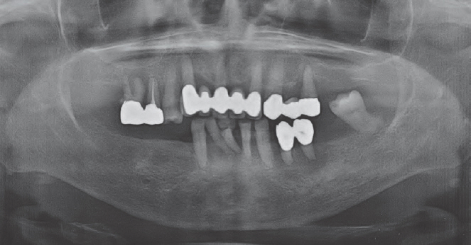
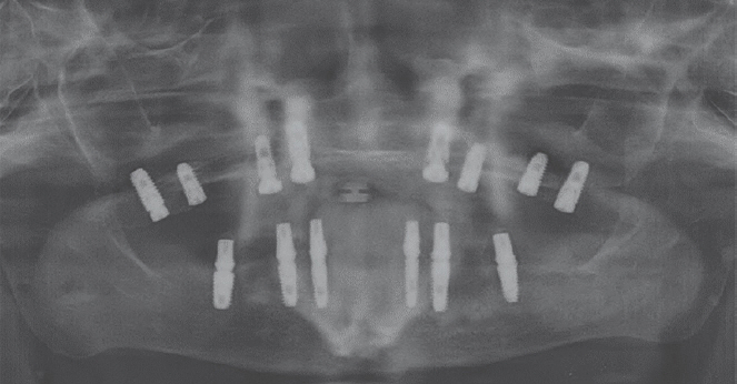
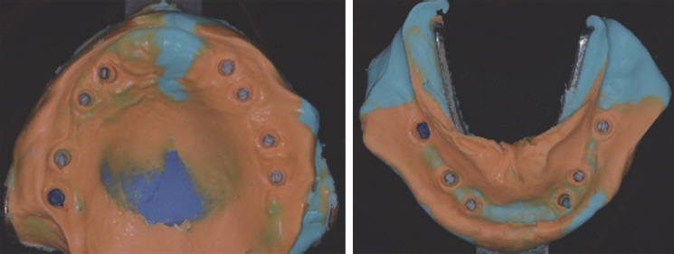
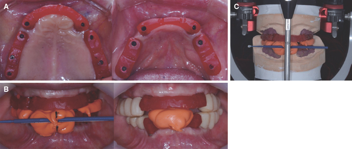
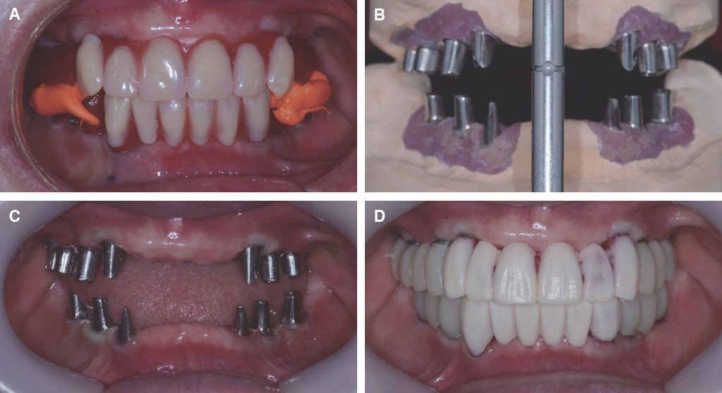
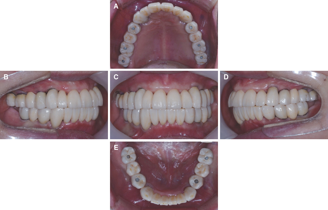
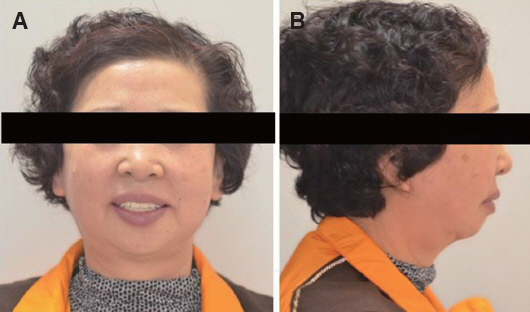
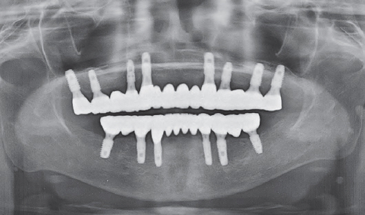
- Oral rehabilitation of a patient having severe periodontitis with alveolar bone resorption and periodontal inflammation presents a challenge to clinicians. However, if appropriate implant placement according to the bone shape is selected, unnecessary bone grafting or soft tissue surgery can be minimized. In recent years, using cone beam CT and software, it has become possible to operate the planned position with the surgical guide made with 3D printing technology. This case was a 70 years old female patient who required total extraction of teeth due to severe periodontitis and performed a full-mouth rehabilitation with an implant-supported fixed prosthesis. During the surgery, the implant was placed in a flapless manner through a surgical guide. Immediate loading of the temporary prosthesis made by CAD/CAM method before surgery was done. Since then, we have produced customized abutments and zirconia prostheses, and have reported satisfactory aesthetic and functional recovery.
MeSH Terms
Figure
Reference
-
References
1. Mankoo T, Frost L. Rehabilitation of esthetics in advanced periodontal cases using orthodontics for vertical hard and soft tissue regeneration prior to implants - a report of 2 challenging cases treated with an interdisciplinary approach. Eur J Esthet Dent. 2011; 6:376–404. PMID: 22238723.2. Chiapasco M, Zaniboni M, Boisco M. Augmentation procedures for the rehabilitation of deficient edentulous ridges with oral implants. Clin Oral Implants Res. 2006; 17(Suppl2):136–59. DOI: 10.1111/j.1600-0501.2006.01357.x. PMID: 16968389.3. Kfir E, Kfir V, Kaluski E. Immediate bone augmentation after infected tooth extraction using titanium membranes. J Oral Implantol. 2007; 33:133–8. DOI: 10.1563/1548-1336(2007)33[133:IBAAIT]2.0.CO;2.4. Jung RE, Schneider D, Ganeles J, Wismeijer D, Zwahlen M, Hämmerle CH, Tahmaseb A. Computer technology applications in surgical implant dentistry: a systematic review. Int J Oral Maxillofac Implants. 2009; 24(Suppl):92–109. PMID: 19885437.5. Hartlev J, Kohberg P, Ahlmann S, Gotfredsen E, Andersen NT, Isidor F, Schou S. Immediate placement and provisionalization of single-tooth implants involving a definitive individual abutment: a clinical and radiographic retrospective study. Clin Oral Implants Res. 2013; 24:652–8. DOI: 10.1111/j.1600-0501.2012.02442.x. PMID: 22409733.6. Schropp L, Isidor F. Timing of implant placement relative to tooth extraction. J Oral Rehabil. 2008; 35(Suppl1):33–43. DOI: 10.1111/j.1365-2842.2007.01827.x. PMID: 18181932.7. Esposito MA, Koukoulopoulou A, Coulthard P, Worthington HV. Interventions for replacing missing teeth: dental implants in fresh extraction sockets (immediate, immediate-delayed and delayed implants). Cochrane Database Syst Rev. 2006; 4:CD005968. DOI: 10.1002/14651858.CD005968.pub2.8. Fortin T, Bosson JL, Isidori M, Blanchet E. Effect of flapless surgery on pain experienced in implant placement using an image-guided system. Int J Oral Maxillofac Implants. 2006; 21:298–304. PMID: 16634502.9. Nkenke E, Eitner S, Radespiel-Tröger M, Vairaktaris E, Neukam FW, Fenner M. Patient-centered outcomes comparing transmucosal implant placement with an open approach in the maxilla: a prospective, non-randomized pilot study. Clin Oral Implants Res. 2007; 18:197–203. DOI: 10.1111/j.1600-0501.2006.01335.x. PMID: 17348884.10. Robling AG, Turner CH. Mechanical signaling for bone modeling and remodeling. Crit Rev Eukaryot Gene Expr. 2009; 19:319–38. DOI: 10.1615/CritRevEukarGeneExpr.v19.i4.50.11. Arisan V, Karabuda CZ, Mumcu E, Özdemir T. Implant positioning errors in freehand and computer-aided placement methods: a single-blind clinical comparative study. Int J Oral Maxillofac Implants. 2013; 28:190–204. DOI: 10.11607/jomi.2691. PMID: 23377066.12. D'haese J, Van De Velde T, Komiyama A, Hultin M, De Bruyn H. Accuracy and complications using computer-designed stereolithographic surgical guides for oral rehabilitation by means of dental implants: a review of the literature. Clin Implant Dent Relat Res. 2012; 14:321–35. DOI: 10.1111/j.1708-8208.2010.00275.x. PMID: 20491822.13. Ozan O, Turkyilmaz I, Ersoy AE, McGlumphy EA, Rosenstiel SF. Clinical accuracy of 3 different types of computed tomography-derived stereolithographic surgical guides in implant placement. J Oral Maxillofacial Surg. 2009; 67:394–401. DOI: 10.1016/j.joms.2008.09.033. PMID: 19138616.14. Di Giacomo GA, Cury PR, de Araujo NS, Sendyk WR, Sendyk CL. Clinical application of stereolithographic surgical guides for implant placement: preliminary results. J Periodontol. 2005; 76:503–7. DOI: 10.1902/jop.2005.76.4.503. PMID: 15857088.15. Papaspyridakos P, Lal K. Complete arch implant rehabilitation using subtractive rapid prototyping and porcelain fused to zirconia prosthesis: a clinical report. J Prosthet Dent. 2008; 100:165–72. DOI: 10.1016/S0022-3913(08)00110-8.16. Kanat B, Cömlekoğlu EM, Dündar-Çömlekoğlu M, Hakan Sen B, Ozcan M, Ali Güngör M. Effect of various veneering techniques on mechanical strength of computer-controlled zirconia framework designs. J Prosthodont. 2014; 23:445–55. DOI: 10.1111/jopr.12130. PMID: 24417370.17. Heintze SD, Rousson V. Survival of zirconia- and metal-supported fixed dental prostheses: a systematic review. Int J Prosthodont. 2010; 23:493–502. PMID: 21209982.
- Full Text Links
- Actions
-
Cited
- CITED
-
- Close
- Share
- Similar articles
-
- Implant-supported fixed prosthesis restoration of fully edentulous patient using computer-guided implant surgery and immediate loading: A case report
- Full mouth rehabilitation in a patient with peri-implantitis: A case report
- Full-mouth rehabilitation of a patient with loss of posterior support and collapsed occlusion utilizing dental CAD-CAM system
- Full mouth rehabilitation with Implant-Guided Surgery and Fixed prosthesis
- Full mouth rehabilitation of a patient using monolithic zirconia and dental CAD/CAM system: a case report


