Full mouth rehabilitation of a patient using monolithic zirconia and dental CAD/CAM system: a case report
- Affiliations
-
- 1Department of Prosthodontics, School of Dentistry, Seoul National University, Seoul, Republic of Korea. ksh1250@snu.ac.kr
- KMID: 2422638
- DOI: http://doi.org/10.14368/jdras.2018.34.3.196
Abstract
- An accurate implant placement with ideal location is significant for long-term success of the implant. An exact evaluation of nearby anatomic structures such as quality of residual bone, an inferior alveolar bone and a maxillary sinus is required. For a prosthetic-driven treatment, planned surgery, precise prosthesis and communication with the patient are significant requisites especially for full-mouth rehabilitation. In this case, the patient with severe alveolar bone resorption had a CT guided surgery supported by CT data and the data from scanning diagnostic wax-up. Afterward, edentulous area was restored by full mouth implant-supported prosthesis by using monolithic zirconia and CAD/CAM technique. This paper reports the outcome of the procedure which was remarkable both esthetically and functionally.
Keyword
MeSH Terms
Figure
-

Fig. 1 Initial panoramic radiographic image. Generalized chronic periodontitis was shown.
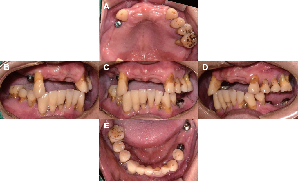
Fig. 2 Intraoral photograph in the initial examination. Generalized chronic periodontitis, implant fixture thread exposure, and irregular occlusal plane was shown. (A) Maxillary occlusal view, (B) Right lateral view, (C) Frontal view at maximum inter cuspal position, (D) Left lateral view, (E) Mandibular occlusal view.
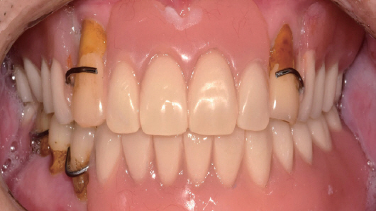
Fig. 3 Temporary denture delivery after teeth extraction.

Fig. 4 Diagnostic wax-up model. (A) Right lateral view, (B) Frontal view, (C) Left lateral view.
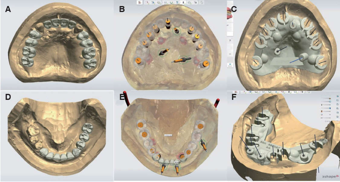
Fig. 5 Fabrication of surgical guide using CAD program. (A) Digital wax up of upper part, (B) Implant planning of upper part, (C) Surgical guide design of upper part, (D) Digital wax up of lower part, (E) Implant planning of lower part, (F) Surgical guide design of lower part.
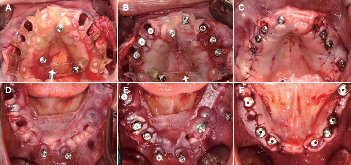
Fig. 6 Implant fixture placement. (A) Adaptation of maxillary surgical guide, (B) Maxilla after implant fixture placement, (C) Maxilla after surgery, (D) Adaptation of mandibular surgical guide, (E) Mandible after implant fixture placement, (F) Mandible after surgery.

Fig. 7 Panoramic radiograph after implant fixture placement.
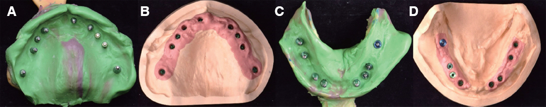
Fig. 8 Implant fixture level impression and master cast fabrication for provisional prosthesis. (A) Pick up coping impression of maxilla, (B) Master cast of maxilla, (C) Pick up coping impression of mandible, (D) Master cast of mandible.

Fig. 9 Vertical dimension determination, facebow transfer, jaw relation registration for provisional prosthesis. (A) Vertical dimension at lateral view, (B) Vertical dimension at frontal view, (C) Facebow transfer, (D) Jaw relation registration.

Fig. 10 Design of customized titanium abutment, digital wax up for provisional prosthesis. (A) Right lateral view, (B) Frontal view, (C) Left lateral view.
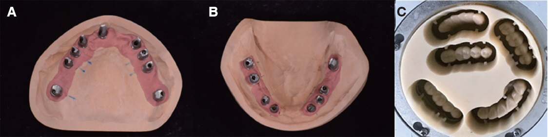
Fig. 11 Fabrication of customized abutment and provisional prosthesis. (A) Customized abutment of maxilla, (B) Customized abutment of mandible, (C) PMMA resin milling.

Fig. 12 Provisional prosthesis. (A) Right lateral view, (B) Frontal view, (C) Left lateral view.

Fig. 13 Final impression and master cast fabrication for definitive prosthesis. (A) Final impression of maxilla, (B) Master cast of maxilla, (C)Final impression of mandible, (D) Master cast of mandible.
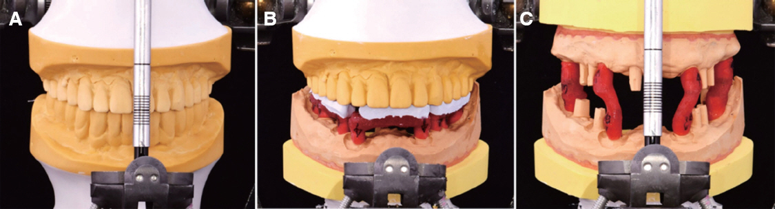
Fig. 14 Cross mounting. (A) Mounting of provisional restoration, (B) Mounting of mandibular master cast, (C) Mounting of maxillary Master cast.

Fig. 15 Secondary provisional prosthesis. Facial profile, lip support, occlusion was re-evaluated.
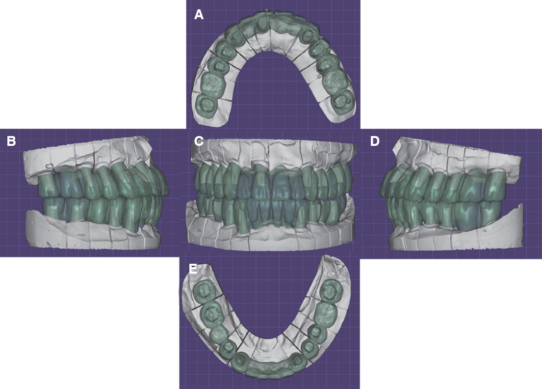
Fig. 16 Digital wax up of definitive prosthesis. (A) Maxillary occlusal view, (B) Right lateral view, (C) Frontal view, (D) Left lateral view, (E) Mandibular occlusal view.
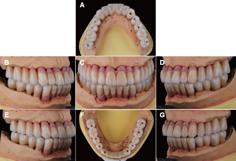
Fig. 17 Definitive prosthesis on master cast. (A) Maxillary occlusal view, (B) Right lateral view, (C) Frontal view, (D) Left lateral view, (E) Lateral movement - right side: group function, (F) Mandibular occlusal view, (G) Lateral movement – left side: group function.
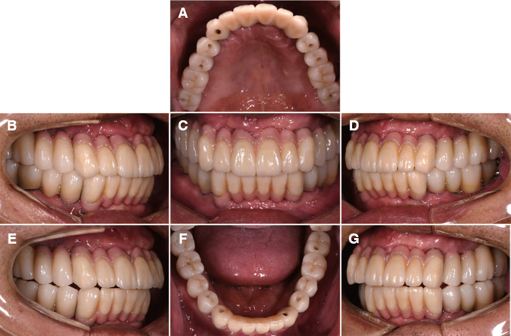
Fig. 18 Definitive prosthesis delivery. Group function with shallow anterior guidance was adopted for occlusal scheme. (A) Maxillary occlusal view, (B) Right lateral view, (C) Frontal view, (D) Left lateral view, (E) Lateral movement – right side: group function, (F) Mandibular occlusal view, (G) Lateral movement - left side: group function.
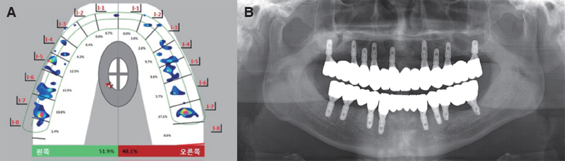
Fig. 19 Definitive prosthesis evaluation. (A) Occlusal analysis using T-scan III: Equal distribution of occlusal force for whole dentition, (B) Post-treatment panoramic radiograph.
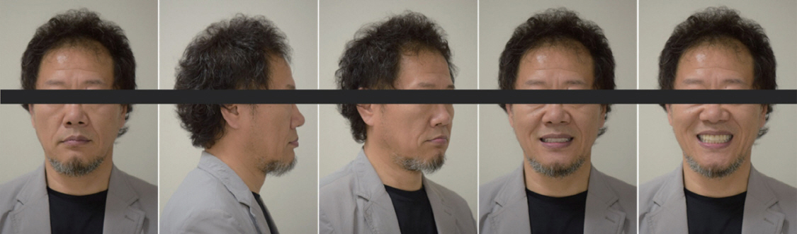
Fig. 20 Extraoral photograph after treatment. The treatment was remarkable both esthetically and functionally.
Reference
-
References
1. Misch CE. Dental implant prosthetics. 2nd ed. St. Louis: Mosby;2015. p. 193–205. DOI: 10.1016/B978-0-323-07845-0.00009-9.2. BouSerhal C, Jacobs R, Quirynen M, van Steenberghe D. Imaging technique selection for the preoperative planning of oral implants:a review of the literature. Clin Implant Dent Relat Res. 2002; 4:156–72. DOI: 10.1111/j.1708-8208.2002.tb00167.x. PMID: 12516649.3. Harris BT, Chen L, Lin WS. Digital Imaging and Prosthetic-Driven Implant Planning:Efficient, Accurate, and Reliable Treatment. Compend Contin Educ Dent. 2017; 38:492–4. PMID: 28727467.4. Ueda K, Güth JF, Erdelt K, Stimmelmayr M, Kappert H, Beuer F. Light transmittance by a multi-coloured zirconia material. Dent Mater J. 2015; 34:310–4. DOI: 10.4012/dmj.2014-238. PMID: 25904173.5. Baldissara P, Wandscher VF, Marchionatti AME, Parisi C, Monaco C, Ciocca L. Translucency of IPS e.max and cubic zirconia monolithic crowns. J Prosthet Dent. 2018; 120:269–75. DOI: 10.1016/j.prosdent.2017.09.007. PMID: 29475752.6. Zitzmann NU, Marinello CP. Treatment plan for restoring the edentulous maxilla with implantsupported restoration:removable overdenture versus fixed partial denture design. J Prosthet Dent. 1999; 82:188–96. DOI: 10.1016/S0022-3913(99)70155-1.7. Raico Gallardo YN, da Silva-Olivio IRT, Mukai E, Morimoto S, Sesma N, Cordaro L. Accuracy comparison of guided surgery for dental implants according to the tissue of support:a systematic review and meta-analysis. Clin Oral Implants Res. 2017; 28:602–12. DOI: 10.1111/clr.12841. PMID: 27062555.8. Sun Y, Luebbers HT, Agbaje JO, Schepers S, Politis C, Van Slycke S, Vrielinck L. Accuracy of Dental Implant Placement Using CBCT-Derived Mucosa-Supported Stereolithographic Template. Clin Implant Dent Relat Res. 2015; 17:862–70. DOI: 10.1111/cid.12189. PMID: 24341829.9. Kim Y, Oh TJ, Misch CE, Wang HL. Occlusal considerations in implant therapy:clinical guidelines with biomechanical rationale. Clin Oral Implants Res. 2005; 16:26–35. DOI: 10.1111/j.1600-0501.2004.01067.x. PMID: 15642028.10. Stawarczyk B, Keul C, Eichberger M, Figge D, Edelhoff D, Lümkemann N. Quintessence Int. 2017; 48:441–50. DOI: 10.3290/j.qi.a38157. PMID: 28497132.11. Wong CKK, Narvekar U, Petridis H. Prosthodontic Complications of Metal-Ceramic and All-Ceramic, Complete-Arch Fixed Implant Prostheses with Minimum 5 Years Mean Follow-Up Period. A Systematic Review and Meta-Analysis. J Prosthodont. 2018; Apr. 17. doi:10.1111/jopr.12797. [Epub ahead of print]. DOI: 10.1111/jopr.12797. PMID: 29665177.12. Moráguez OD, Wiskott HW, Scherrer SS. Three- to nine-year survival estimates and fracture mechanisms of zirconia- and alumina-based restorations using standardized criteria to distinguish the severity of ceramic fractures. Clin Oral Investig. 2015; 19:2295–307. DOI: 10.1007/s00784-015-1455-y. PMID: 25986462.13. Bidra AS, Rungruanganunt P, Gauthier M. Clinical outcomes of full arch fixed implant-supported zirconia prostheses:a systematic review. Eur J Oral Implantol. 2017; 10:35–45. PMID: 28944367.
- Full Text Links
-
- Actions
-
Cited
- CITED
-
- Close
- Share
-
- Similar articles
-
- Full mouth rehabilitation using monolithic zirconia: a clinical report
- Maxillary cement retained implant supported monolithic zirconia prosthesis in a full mouth rehabilitation: a clinical report
- A full-mouth rehabilitation using zirconia all-ceramic crowns: a case report
- Full mouth rehabilitation with vertical dimension increase in patient with severly worn dentition
- Full mouth rehabilitation in a patient with partial mandibulectomy using CAD/CAM zirconia framework and monolithic zirconia
