A full-mouth rehabilitation using zirconia all-ceramic crowns: a case report
- Affiliations
-
- 1Department of Prosthodontics, School of Dentistry and Institute of Oral Bio-Science, Chonbuk National University, Jeonju, Republic of Korea. jmseo@jbnu.ac.kr
- 2Department of Dentistry, School of Medicine, Eulji University, Daejeon, Republic of Korea.
- KMID: 2180024
- DOI: http://doi.org/10.14368/jdras.2015.31.3.231
Abstract
- As patients' esthetic expectations increase, there is an increase in demand for cosmetic restorations of anterior and posterior teeth that resemble the natural tooth morphology and color. An example of high-strength all-ceramic systems is zirconia with CAD/CAM application. This case report describes a full-mouth rehabilitation using zirconia all-ceramic crowns supported by upper and lower implants and by a minimal increase in the occlusal vertical dimension in a patient with severe abrasion due to loss of posterior teeth.
Keyword
Figure
-

Fig. 1 Intra-oral status in the initial examination. (A) Maxillary occlusal view, (B) Right lateral view, (C) Frontal view at maximum inter-cuspal position (D) Left lateral view, (E) Mandibular occlusal view.

Fig. 2 Pre-treatment panoramic radiograph.

Fig. 3 Diagnostic wax-up model with increased vertical dimension. (A) Maxillary occlusal view, (B) Right lateral view, (C) Frontal view, (D) Left lateral view, (E) Mandibular occlusal view.

Fig. 4 Occlusion of eccentric movements. (A) Anterior movement, (B) Lateral movement- right side: group function, (C) Lateral movement- left side: canine guidance.
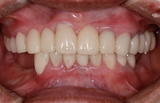
Fig. 5 1st provisional restoration.

Fig. 6 Reproduction of the existing gingival contour with a putty for customized impression coping of #12, 13 implant.

Fig. 7 Working cast for definitive prosthesis. (A) Maxillary occlusal view, (B) Mandibular occlusal view.

Fig. 8 Registration of inter-occlusal relationship with the provisional restoration.
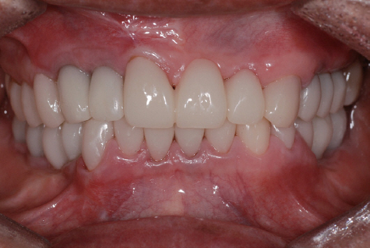
Fig. 9 Frontal view of the new provisional restoration.

Fig. 10 Customized abutment for implant. (A) Maxillary occlusal view, (B) Mandibular occlusal view.

Fig. 11 Full contour wax-up for the definitive prosthesis. (A) Maxillary occlusal view, (B) Right lateral view, (C) Frontal view at maximum inter-cuspal position (D) Left lateral view, (E) Mandibular occlusal view.
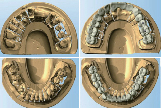
Fig. 12 Double scan images of working cast and wax-up model were superimposed.
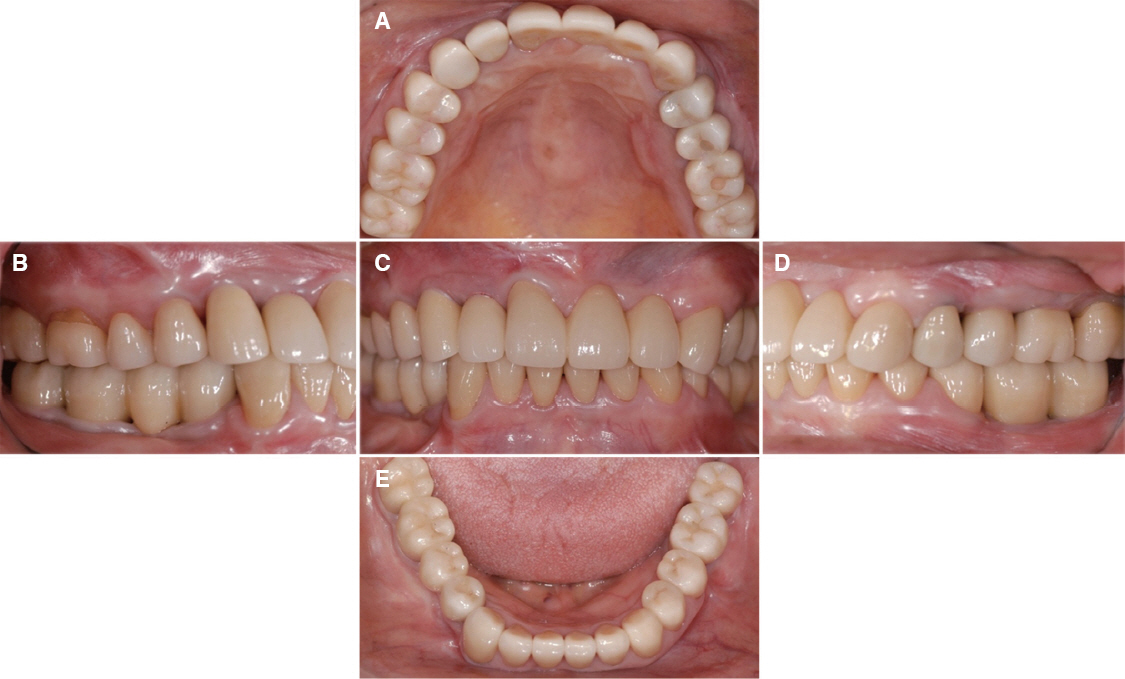
Fig. 13 Definitive prosthesis was delivered. Esthetics and functions were restored with the zirconia prosthesis. (A) Maxillary occlusal view, (B) Right lateral view, (C) Frontal view at maximum inter-cuspal position, (D) Left lateral view, (E) Mandibular occlusal view.
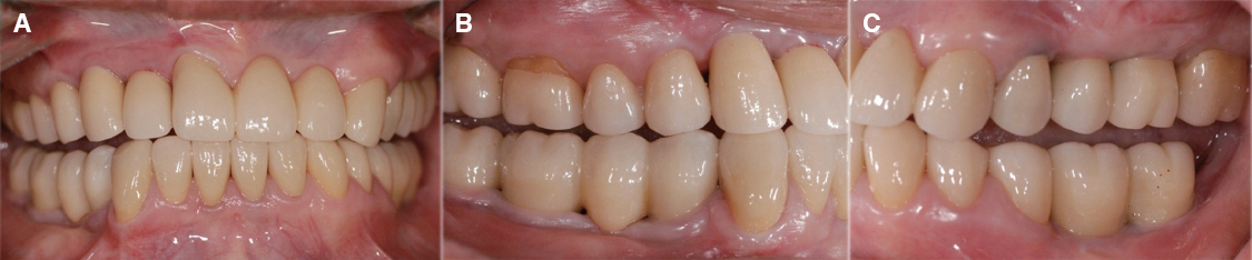
Fig. 14 Eccentric occlusion with definitive prosthesis. (A) Anterior movement: posterior teeth of both side were disoccluded, (B) Lateral movement - right side: group function, (C) Lateral movement - left side: canine guidance.
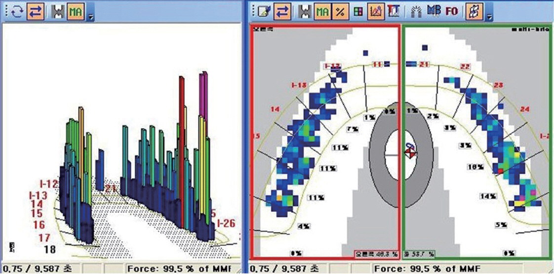
Fig. 15 Occlusal analysis using T scan III. Stable occlusion was present.
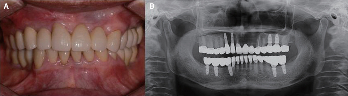
Fig. 16 Follow-up check after 3 years. (A) Stable occlusion and good oral hygiene were maintained, (B) Post-treatment panoramic radiograph.
Reference
-
References
1. Anusavice KJ. Recent developments in restorative dental ceramics. J Am Dent Assoc. 1993; 124:72–4. 76-8, 80-4. DOI: 10.14219/jada.archive.1993.0031. PMID: 8429187.2. Bagby M, Marshall SJ, Marshall GW Jr. Metal ceramic compatibility: a review of the literature. J Prosthet Dent. 1990; 63:21–5. DOI: 10.1016/0022-3913(90)90259-F.3. Campbell SD. A comparative strength study of metal ceramic and all-ceramic esthetic materials: modulus of rupture. J Prosthet Dent. 1989; 62:4769. DOI: 10.1016/0022-3913(89)90184-4.4. Kelly JR. Dental ceramics: current thinking and trends. Dent Clin North Am. 2004; 48:513–30. DOI: 10.1016/j.cden.2004.01.003. PMID: 15172614.5. McLaren EA. All-ceramic alternatives to conventional metal-ceramic restorations. Compend Contin Educ Dent. 1998; 19:307–8. 310, 312 passim; quiz 326.6. Guess PC, Schultheis S, Bonfante EA, Coelho PG, Ferencz JL, Silva NR. All-ceramic systems: laboratory and clinical performance. Dent Clin North Am. 2011; 55:333–52. DOI: 10.1016/j.cden.2011.01.005. PMID: 21473997.7. Vult von Steyern P, Carlson P, Nilner K. All-ceramic fixed partial dentures designed according to the DC-Zirkon technique. A 2-year clinical study. J Oral Rehabil. 2005; 32:180–7. DOI: 10.1111/j.1365-2842.2004.01437.x. PMID: 15707428.8. Bouri A, Bissada N, Al-Zahrani MS, Faddoul F, Nouneh I. Width of keratinized gingiva and the health status of the supporting tissues around dental implants. Int J Oral Maxillofac Implants. 2008; 23:323–6. PMID: 18548930.9. Zigdon H, Machtei EE. The dimensions of keratinized mucosa around implants affect clinical and immunological parameters. Clin Oral Implants Res. 2008; 19:387–92. DOI: 10.1111/j.1600-0501.2007.01492.x. PMID: 18266873.10. Wood GN. Centric relation and the treatment position in rehabilitating occlusions: a physiologic approach. Part II: The treatment position. J Prosthet Dent. 1988; 60:15–8. DOI: 10.1016/0022-3913(88)90341-1.11. Fayz F, Eslami A. Determination of occlusal vertical dimension: a literature review. J Prosthet Dent. 1988; 59:321–3. DOI: 10.1016/0022-3913(88)90182-5.12. Abduo J. Safety of increasing vertical dimension of occlusion: a systematic review. Quintessence int. 2012; 43:369–80. PMID: 22536588.13. Ozkurt Z, Kazazoğlu E. Clinical success of zirconia in dental applications. J Prosthodont. 2010; 19:64–8. DOI: 10.1111/j.1532-849X.2009.00513.x. PMID: 19754642.14. Bachhav VC, Aras MA. Zirconia-based fixed partial dentures: a clinical review. Quintessence Int. 2011; 42:173–82. PMID: 21359252.15. Al-Amleh B, Lyons K, Swain M. Clinical trials in zirconia: a systematic review. J Oral Rehabil. 2010; 37:641–52. DOI: 10.1111/j.1365-2842.2010.02094.x.16. Choi BK, Han JS, Yang JH, Lee JB, Kim SH. Shear bond strength of veneering porcelain to zirconia and metal cores. J Adv Prosthodont. 2009; 1:129–35. DOI: 10.4047/jap.2009.1.3.129. PMID: 21165268. PMCID: PMC2994690.
- Full Text Links
-
- Actions
-
Cited
- CITED
-
- Close
- Share
-
- Similar articles
-
- Full mouth rehabilitation using monolithic zirconia: a clinical report
- Marginal fit of the digident CAD/CAM zirconia ceramic crowns
- Full mouth rehabilitation using zirconia crown in severe worn dentition: a case report
- Fracture strength of zirconia ceramic crowns according to tooth position
- Full mouth rehabilitation of a patient with tooth wear and insufficient restorative space due to loss of posterior teeth support: a case report
