Image-Enhanced Endoscopy in Lower Gastrointestinal Diseases: Present and Future
- Affiliations
-
- 1Division of Gastroenterology, Department of Internal Medicine, College of Medicine, The Catholic University of Korea, Seoul, Korea. gidoc4u@gmail.com
- 2Catholic Photomedicine Research Institute, Seoul, Korea.
- KMID: 2430394
- DOI: http://doi.org/10.5946/ce.2018.187
Abstract
- From dye-assisted conventional chromoendoscopy to novel virtual chromoendoscopy, image-enhanced endoscopy (IEE) is continuously evolving to meet clinical needs and improve the quality of colonoscopy. Dye-assisted chromoendoscopy using indigo carmine or crystal violet, although slightly old-fashioned, is still useful to emphasize the pit patterns of the colonic mucosa and predict the histological structures of relevant lesions. Equipment-based virtual chromoendoscopy has the advantage of being relatively easy to use. There are several types of virtual chromoendoscopy that vary depending on the manufacturer and operating principle. IEE plays distinctive roles with respect to histologic characterization of colorectal polyps and prediction of the invasion depth of colorectal cancers. In addition, the newest models of IEE have the potential to increase adenoma and polyp detection rates in screening colonoscopy.
MeSH Terms
Figure
-
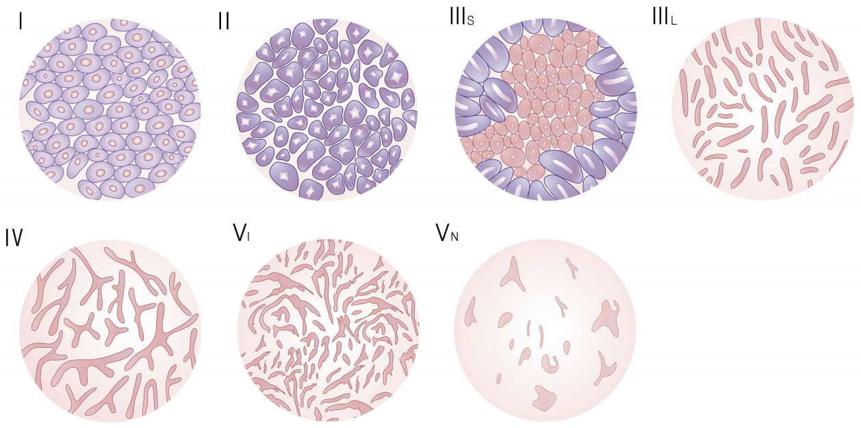
Fig. 1. Schematic presentation of Kudo’s pit-pattern classification.
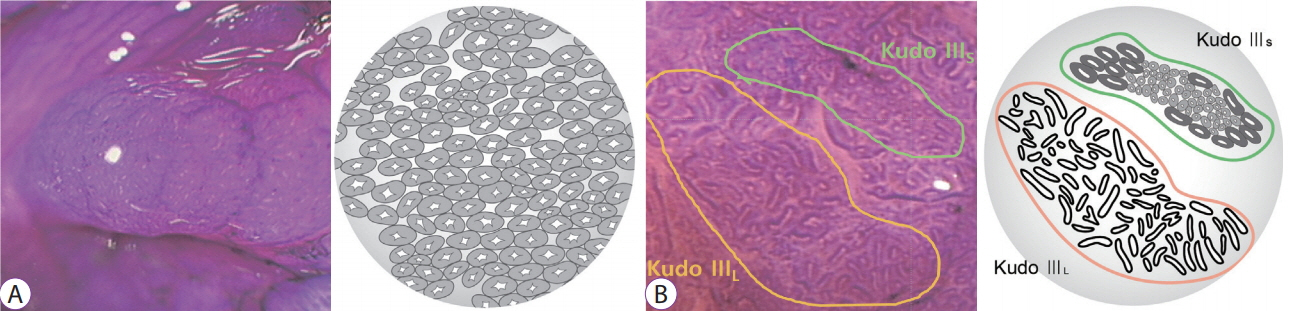
Fig. 2. Prediction of the histological structure of the detected lesion by magnifying chromoendoscopy using crystal violet. (A) The pits of the polyp appear to be pressed flat, and they are regularly arranged. This pit-pattern is similar to Kudo’s type II pattern, which suggests a hyperplastic polyp. (B) The pit-pattern of the lesion shows a mixed pattern of Kudo’s types IIIL and IIIs. This lesion was confirmed as being an intraepithelial adenocarcinoma.
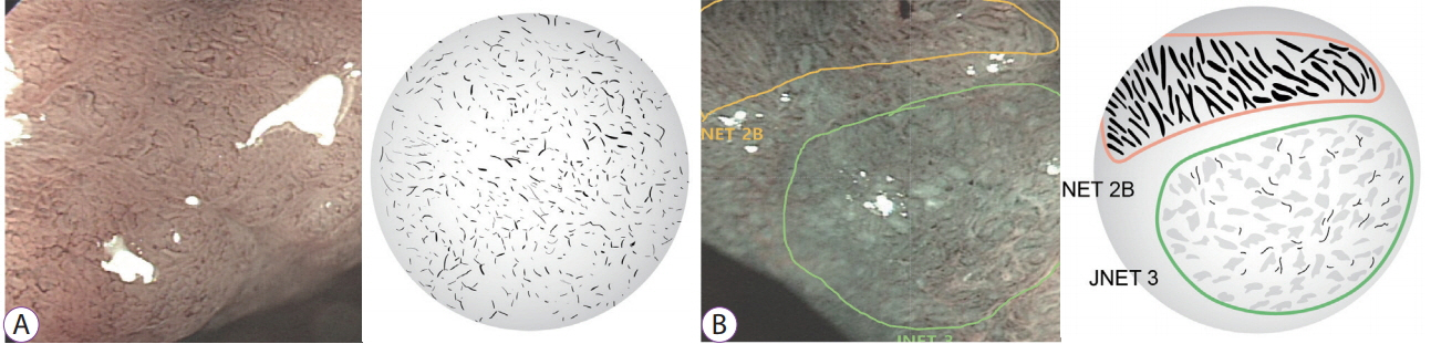
Fig. 3. Prediction of the invasion depth of the detected lesion by magnifying narrow-band imaging. (A) The lesion shows a capillary pattern characterized by a blind ending, lack of uniformity, and curtailed irregularly, which is similar to the Sano IIIA pattern. This lesion was confirmed as being an intraepithelial adenocarcinoma. (B) The slightly depressed center of the lesion shows loose vessel areas and an amorphous surface pattern. This lesion was confirmed as being a deep submucosal invasive adenocarcinoma.
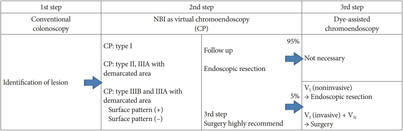
Fig. 4. Three-step strategy of narrow-band imaging (NBI) colonoscopy. CP, capillary pattern.
Reference
-
1. Kudo S, Rubio CA, Teixeira CR, Kashida H, Kogure E. Pit pattern in colorectal neoplasia: endoscopic magnifying view. Endoscopy. 2001; 33:367–373.
Article2. Muto M, Horimatsu T, Ezoe Y, Morita S, Miyamoto S. Improving visualization techniques by narrow band imaging and magnification endoscopy. J Gastroenterol Hepatol. 2009; 24:1333–1346.
Article3. Gono K, Obi T, Yamaguchi M, et al. Appearance of enhanced tissue features in narrow-band endoscopic imaging. J Biomed Opt. 2004; 9:568–577.
Article4. Sano Y, Ikematsu H, Fu KI, et al. Meshed capillary vessels by use of narrow-band imaging for differential diagnosis of small colorectal polyps. Gastrointest Endosc. 2009; 69:278–283.
Article5. Katagiri A, Fu KI, Sano Y, et al. Narrow band imaging with magnifying colonoscopy as diagnostic tool for predicting histology of early colorectal neoplasia. Aliment Pharmacol Ther. 2008; 27:1269–1274.
Article6. Ikematsu H, Matsuda T, Emura F, et al. Efficacy of capillary pattern type IIIA/IIIB by magnifying narrow band imaging for estimating depth of invasion of early colorectal neoplasms. BMC Gastroenterol. 2010; 10:33.
Article7. Kanao H, Tanaka S, Oka S, Hirata M, Yoshida S, Chayama K. Narrow-band imaging magnification predicts the histology and invasion depth of colorectal tumors. Gastrointest Endosc. 2009; 69(3 Pt 2):631–636.
Article8. Wada Y, Kudo SE, Kashida H, et al. Diagnosis of colorectal lesions with the magnifying narrow-band imaging system. Gastrointest Endosc. 2009; 70:522–531.
Article9. Hewett DG, Kaltenbach T, Sano Y, et al. Validation of a simple classification system for endoscopic diagnosis of small colorectal polyps using narrow-band imaging. Gastroenterology. 2012; 143:599–607. e1.
Article10. Hayashi N, Tanaka S, Hewett DG, et al. Endoscopic prediction of deep submucosal invasive carcinoma: validation of the narrow-band imaging international colorectal endoscopic (NICE) classification. Gastrointest Endosc. 2013; 78:625–632.
Article11. Sano Y, Tanaka S, Kudo SE, et al. Narrow-band imaging (NBI) magnifying endoscopic classification of colorectal tumors proposed by the Japan NBI expert team. Dig Endosc. 2016; 28:526–533.
Article12. Pohl J, Lotterer E, Balzer C, et al. Computed virtual chromoendoscopy versus standard colonoscopy with targeted indigocarmine chromoscopy: a randomised multicentre trial. Gut. 2009; 58:73–78.
Article13. Aminalai A, Rösch T, Aschenbeck J, et al. Live image processing does not increase adenoma detection rate during colonoscopy: a randomized comparison between FICE and conventional imaging (Berlin colonoscopy project 5, BECOP-5). Am J Gastroenterol. 2010; 105:2383–2388.
Article14. Chung SJ, Kim D, Song JH, et al. Efficacy of computed virtual chromoendoscopy on colorectal cancer screening: a prospective, randomized, back-to-back trial of Fuji intelligent color enhancement versus conventional colonoscopy to compare adenoma miss rates. Gastrointest Endosc. 2010; 72:136–142.
Article15. Osawa H, Yamamoto H. Present and future status of flexible spectral imaging color enhancement and blue laser imaging technology. Dig Endosc. 2014; 26 Suppl 1:105–115.
Article16. Rondonotti E, Paggi S, Amato A, et al. Blue-light imaging compared with high-definition white light for real-time histology prediction of colorectal polyps less than 1 centimeter: a prospective randomized study. Gastrointest Endosc. 2018; Sep. 28. [Epub]. https://doi.org/10.1016/j.gie.2018.09.027.
Article17. Sun X, Dong T, Bi Y, et al. Linked color imaging application for improving the endoscopic diagnosis accuracy: a pilot study. Sci Rep. 2016; 6:33473.
Article18. Hoffman A, Sar F, Goetz M, et al. High definition colonoscopy combined with i-Scan is superior in the detection of colorectal neoplasias compared with standard video colonoscopy: a prospective randomized controlled trial. Endoscopy. 2010; 42:827–833.
Article19. Hong SN, Choe WH, Lee JH, et al. Prospective, randomized, back-toback trial evaluating the usefulness of i-Scan in screening colonoscopy. Gastrointest Endosc. 2012; 75:1011–1021. e2.
Article20. Brown SR, Baraza W, Din S, Riley S. Chromoscopy versus conventional endoscopy for the detection of polyps in the colon and rectum. Cochrane Database Syst Rev. 2016; 4:CD006439.
Article21. Brooker JC, Saunders BP, Shah SG, et al. Total colonic dye-spray increases the detection of diminutive adenomas during routine colonoscopy: a randomized controlled trial. Gastrointest Endosc. 2002; 56:333–338.
Article22. Jin XF, Chai TH, Shi JW, Yang XC, Sun QY. Meta-analysis for evaluating the accuracy of endoscopy with narrow band imaging in detecting colorectal adenomas. J Gastroenterol Hepatol. 2012; 27:882–887.
Article23. Dinesen L, Chua TJ, Kaffes AJ. Meta-analysis of narrow-band imaging versus conventional colonoscopy for adenoma detection. Gastrointest Endosc. 2012; 75:604–611.
Article24. Nagorni A, Bjelakovic G, Petrovic B. Narrow band imaging versus conventional white light colonoscopy for the detection of colorectal polyps. Cochrane Database Syst Rev. 2012; 1:CD008361.
Article25. Pasha SF, Leighton JA, Das A, et al. Comparison of the yield and miss rate of narrow band imaging and white light endoscopy in patients undergoing screening or surveillance colonoscopy: a meta-analysis. Am J Gastroenterol. 2012; 107:363–370. quiz 371.
Article26. Omata F, Ohde S, Deshpande GA, Kobayashi D, Masuda K, Fukui T. Image-enhanced, chromo, and cap-assisted colonoscopy for improving adenoma/neoplasia detection rate: a systematic review and meta-analysis. Scand J Gastroenterol. 2014; 49:222–237.
Article27. Rey JF, Tanaka S, Lambert R, Tajiri H. Evaluation of the clinical outcomes associated with EXERA II and LUCERA endoscopes. Dig Endosc. 2009; 21 Suppl 1:S113–S120.
Article28. Leung WK, Lo OS, Liu KS, et al. Detection of colorectal adenoma by narrow band imaging (HQ190) vs. high-definition white light colonoscopy: a randomized controlled trial. Am J Gastroenterol. 2014; 109:855–863.
Article29. Yoshida N, Hisabe T, Hirose R, et al. Improvement in the visibility of colorectal polyps by using blue laser imaging (with video). Gastrointest Endosc. 2015; 82:542–549.
Article30. Shimoda R, Sakata Y, Fujise T, et al. The adenoma miss rate of blue-laser imaging vs. white-light imaging during colonoscopy: a randomized tandem trial. Endoscopy. 2017; 49:186–190.
Article31. Suzuki T, Hara T, Kitagawa Y, et al. Linked-color imaging improves endoscopic visibility of colorectal nongranular flat lesions. Gastrointest Endosc. 2017; 86:692–697.
Article32. Wanders LK, East JE, Uitentuis SE, Leeflang MM, Dekker E. Diagnostic performance of narrowed spectrum endoscopy, autofluorescence imaging, and confocal laser endomicroscopy for optical diagnosis of colonic polyps: a meta-analysis. Lancet Oncol. 2013; 14:1337–1347.
Article33. Hassan C, Pickhardt PJ, Rex DK. A resect and discard strategy would improve cost-effectiveness of colorectal cancer screening. Clin Gastroenterol Hepatol. 2010; 8:865–869. e1-e3.
Article34. Yoshida N, Hisabe T, Inada Y, et al. The ability of a novel blue laser imaging system for the diagnosis of invasion depth of colorectal neoplasms. J Gastroenterol. 2014; 49:73–80.
Article35. Nakajima T, Saito Y, Tanaka S, et al. Current status of endoscopic resection strategy for large, early colorectal neoplasia in Japan. Surg Endosc. 2013; 27:3262–3270.
Article36. Matsuda T, Fujii T, Saito Y, et al. Efficacy of the invasive/non-invasive pattern by magnifying chromoendoscopy to estimate the depth of invasion of early colorectal neoplasms. Am J Gastroenterol. 2008; 103:2700–2706.
Article37. Iwatate M, Ikumoto T, Hattori S, Sano W, Sano Y, Fujimori T. NBI and NBI combined with magnifying colonoscopy. Diagn Ther Endosc. 2012; 2012:173269.
Article38. Suzuki T, Hara T, Kitagawa Y, Yamaguchi T. Magnified endoscopic observation of early colorectal cancer by linked color imaging with crystal violet staining (with video). Gastrointest Endosc. 2016; 84:726–729.
Article
- Full Text Links
-
- Actions
-
Cited
- CITED
-
- Close
- Share
-
- Similar articles
-
- Is Image-Enhanced Endoscopy Useful for the Diagnosis and Treatment of Gastrointestinal Tumor?
- The Past, Present, and Future of Image-Enhanced Endoscopy
- Past, Present, and Future of the Korea-Japan Joint Symposium on Gastrointestinal Endoscopy
- Role of Image-Enhanced Endoscopy in Pancreatobiliary Diseases
- Quality Improvement of Gastrointestinal Endoscopy in Korea: Past, Present, and Future
