Clin Endosc.
2018 Nov;51(6):541-546. 10.5946/ce.2018.203.
Role of Image-Enhanced Endoscopy in Pancreatobiliary Diseases
- Affiliations
-
- 1Digestive Disease Center and Research Institute, Department of Internal Medicine, Soonchunhyang University College of Medicine, Bucheon, Korea. jhmoon@schmc.ac.kr
- KMID: 2430395
- DOI: http://doi.org/10.5946/ce.2018.203
Abstract
- Recent advances in cholangiopancreatoscopy technology permit image-enhanced endoscopy (IEE) for pancreatobiliary diseases. There are limitations in endoscopy performance and in the study of the clinical role of IEE in bile duct or pancreatic duct diseases. However, currently available IEEs during cholangiopancreatoscopy including traditional dye-aided chromoendoscopy, autofluorescence imaging, narrow-band imaging, and i-Scan have been evaluated and reported previously. Although the clinical role of IEE in pancreatobiliary diseases should be verified in future studies, IEE is a useful promising tool in the evaluation of bile duct or pancreatic duct mucosal lesions.
MeSH Terms
Figure
-
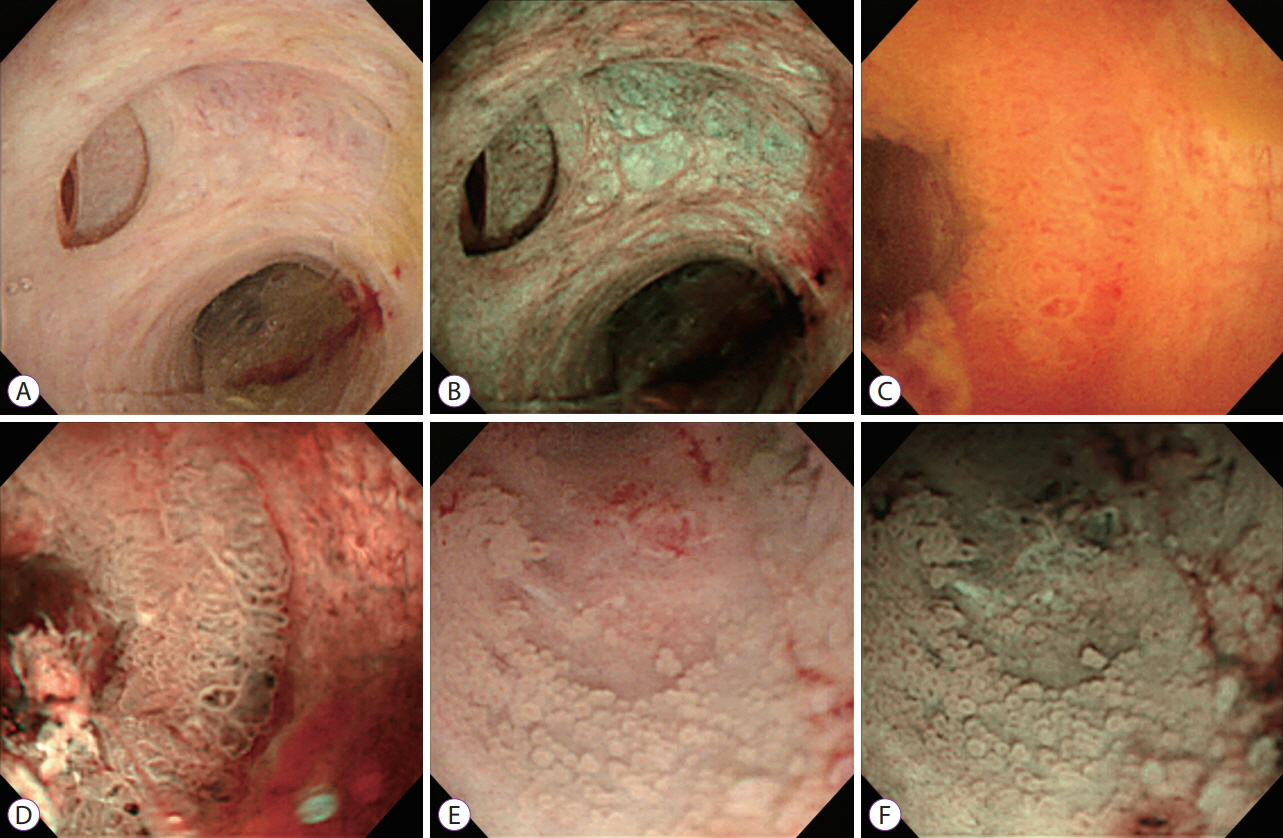
Fig. 1. Peroral cholangioscopy with narrow-band imaging (NBI). White light image (A) and NBI (B) of the normal bile duct. White light image (C and E) and NBI (D and F) of intraductal papillary neoplasm in the bile duct.
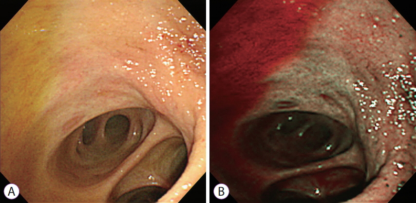
Fig. 2. Peroral cholangioscopy with narrow-band imaging (NBI). Bile appears as red-colored fluid in NBI.
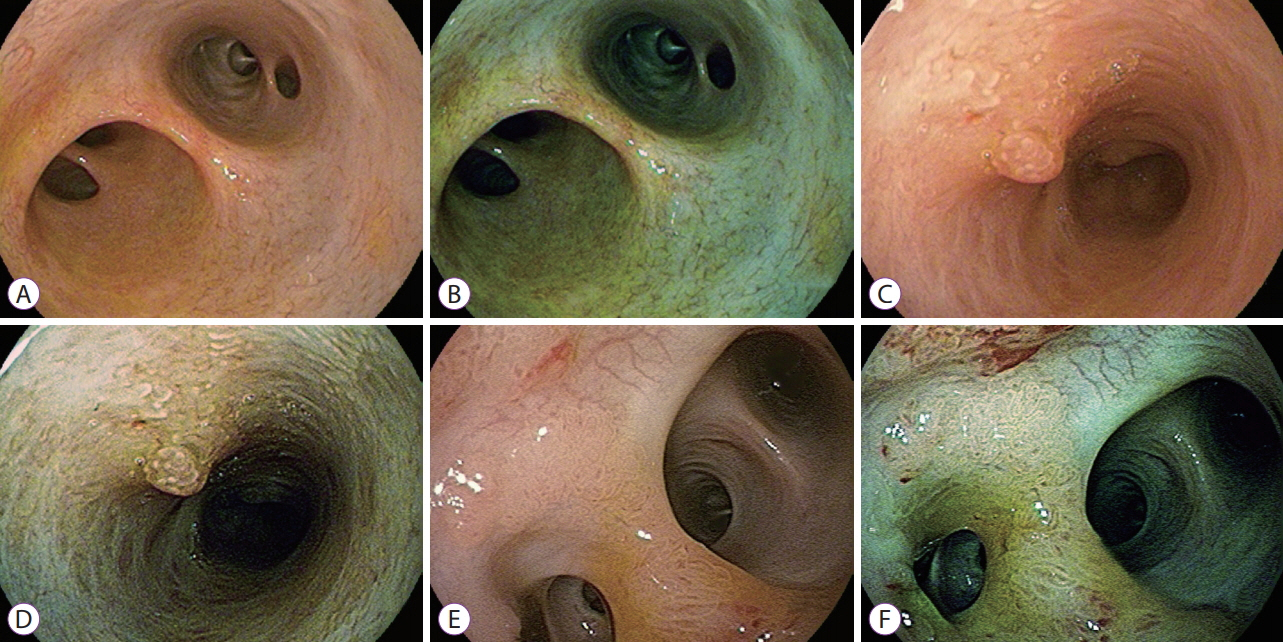
Fig. 3. Peroral cholangioscopy with i-Scan. White light image (A) and i-Scan image (B) of normal bile duct at the hilar bifurcation. White light image (C) and i-Scan image (D) of an inflammatory polyp. White light image (E) and i-Scan image (F) of an intraductal papillary neoplasm in the bile duct.
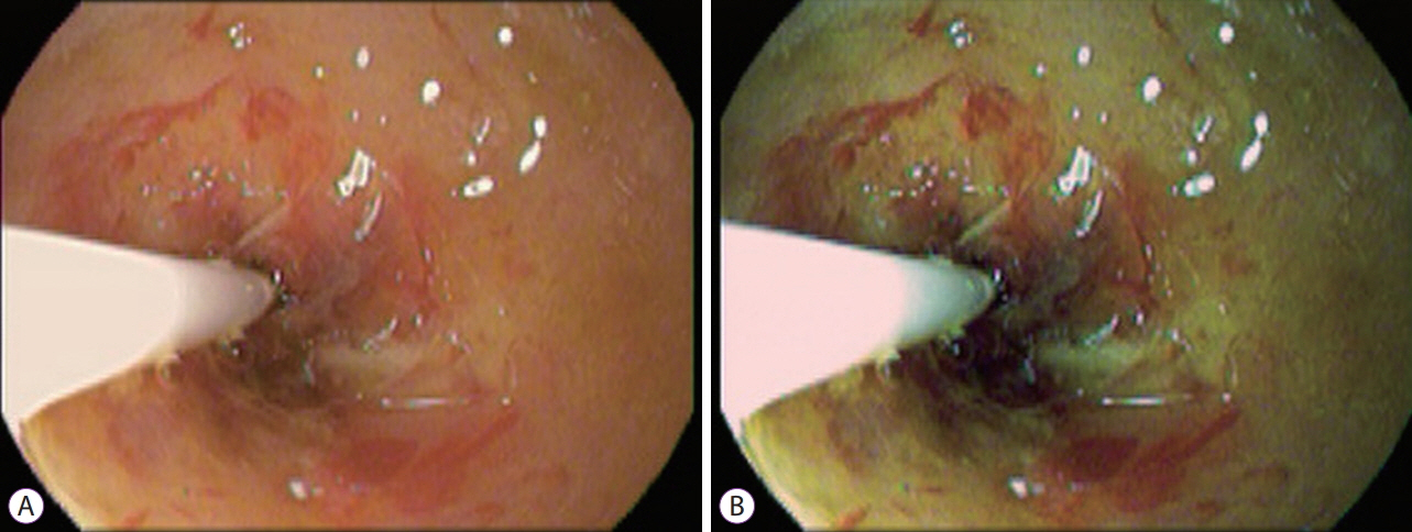
Fig. 4. Twin mode of i-Scan.
Reference
-
1. Moon JH, Terheggen G, Choi HJ, Neuhaus H. Peroral cholangioscopy: diagnostic and therapeutic applications. Gastroenterology. 2013; 144:276–282.
Article2. Kim WJ, Park SY, Park I, et al. Increased detection of colorectal polyps in screening colonoscopy using high definition i-Scan compared with standard white light. Clin Endosc. 2016; 49:69–75.
Article3. Sun X, Zhou Z, Tian J, et al. Is single-operator peroral cholangioscopy a useful tool for the diagnosis of indeterminate biliary lesion? A systematic review and meta-analysis. Gastrointest Endosc. 2015; 82:79–87.
Article4. Lee YN, Moon JH, Choi HJ, et al. Direct peroral cholangioscopy for diagnosis of bile duct lesions using an i-Scan ultraslim endoscope: a pilot study. Endoscopy. 2017; 49:675–681.
Article5. Mounzer R, Austin GL, Wani S, Brauer BC, Fukami N, Shah RJ. Per-oral video cholangiopancreatoscopy with narrow-band imaging for the evaluation of indeterminate pancreaticobiliary disease. Gastrointest Endosc. 2017; 85:509–517.6. Ishida Y, Itoi T, Okabe Y. Can image-enhanced cholangioscopy distinguish benign from malignant lesions in the biliary duct? Best Pract Res Clin Gastroenterol. 2015; 29:611–625.
Article7. Itoi T, Sofuni A, Itokawa F, et al. Initial experience of peroral pancreatoscopy combined with narrow-band imaging in the diagnosis of intraductal papillary mucinous neoplasms of the pancreas (with videos). Gastrointest Endosc. 2007; 66:793–797.
Article8. Itoi T, Sofuni A, Itokawa F, et al. Peroral cholangioscopic diagnosis of biliary-tract diseases by using narrow-band imaging (with videos). Gastrointest Endosc. 2007; 66:730–736.
Article9. Parsi MA, Jang S, Sanaka M, Stevens T, Vargo JJ. Diagnostic and therapeutic cholangiopancreatoscopy: performance of a new digital cholangioscope. Gastrointest Endosc. 2014; 79:936–942.
Article10. Ishida Y, Itoi T, Okabe Y. Types of peroral cholangioscopy: how to choose the most suitable type of cholangioscopy. Curr Treat Options Gastroenterol. 2016; 14:210–219.
Article11. ASGE Technology Committee, Komanduri S, Thosani N, et al. Cholangiopancreatoscopy. Gastrointest Endosc. 2016; 84:209–221.
Article12. Choi JH, Lee SK. Percutaneous transhepatic cholangioscopy: does its role still exist? Clin Endosc. 2013; 46:529–536.
Article13. Hamada T, Tsujino T, Sasahira N, et al. Percutaneous transhepatic cholangioscopy with an ultraslim video upper endoscope with CO2 insufflation: a feasibility study. Gastrointest Endosc. 2011; 74:696–699.
Article14. Maetani I, Ogawa S, Sato M, Igarashi Y, Sakai Y, Shibuya K. Lack of methylene blue staining in superficial epithelia as a possible marker for superficial lateral spread of bile duct cancer. Diagn Ther Endosc. 1996; 3:29–34.
Article15. Hoffman A, Kiesslich R, Bittinger F, Galle PR, Neurath MF. Methylene blue-aided cholangioscopy in patients with biliary strictures: feasibility and outcome analysis. Endoscopy. 2008; 40:563–571.
Article16. Itoi T, Shinohara Y, Takeda K, Nakamura K, Takei K. Improvement of choledochoscopy: chromoendocholedochoscopy, autofluorescence imaging, or narrow-band imaging. Dig Endosc. 2007; 19(Suppl 1):S95–S104.
Article17. Osanai M, Itoi T, Igarashi Y, et al. Peroral video cholangioscopy to evaluate indeterminate bile duct lesions and preoperative mucosal cancerous extension: a prospective multicenter study. Endoscopy. 2013; 45:635–642.
Article18. Nishikawa T, Tsuyuguchi T, Sakai Y, et al. Preoperative assessment of longitudinal extension of cholangiocarcinoma with peroral video-cholangioscopy: a prospective study. Dig Endosc. 2014; 26:450–457.
Article19. Azeem N, Gostout CJ, Knipschield M, Baron TH. Cholangioscopy with narrow-band imaging in patients with primary sclerosing cholangitis undergoing ERCP. Gastrointest Endosc. 2014; 79:773–779. e2.
Article20. Ishida Y, Okabe Y, Kaji R, et al. Evaluation of magnifying endoscopy using narrow band imaging using ex vivo bile duct (with video). Dig Endosc. 2013; 25:322–328.21. Ishida Y, Okabe Y, Yasumoto M, et al. Ex vivo magnifying endoscopic observation of bile duct mucosa using narrowband imaging. J Hepatobiliary Pancreat Sci. 2018; 25:433–439.
Article22. Brauer BC, Fukami N, Chen YK. Direct cholangioscopy with narrow-band imaging, chromoendoscopy, and argon plasma coagulation of intraductal papillary mucinous neoplasm of the bile duct (with videos). Gastrointest Endosc. 2008; 67:574–576.
Article23. Itoi T, Sofuni A, Itokawa F, Tsuchiya T, Kurihara T. Evaluation of peroral videocholangioscopy using narrow-band imaging for diagnosis of intraductal papillary neoplasm of the bile duct. Dig Endosc. 2009; 21 Suppl 1:S103–S107.
Article24. Itoi T, Neuhaus H, Chen YK. Diagnostic value of image-enhanced video cholangiopancreatoscopy. Gastrointest Endosc Clin N Am. 2009; 19:557–566.
Article25. Itoi T, Kamisawa T, Igarashi Y, et al. The role of peroral video cholangioscopy in patients with IgG4-related sclerosing cholangitis. J Gastroenterol. 2013; 48:504–514.
Article
- Full Text Links
-
- Actions
-
Cited
- CITED
-
- Close
- Share
-
- Similar articles
-
- Is Image-Enhanced Endoscopy Useful for the Diagnosis and Treatment of Gastrointestinal Tumor?
- Application of Current Image-Enhanced Endoscopy in Gastric Diseases
- Current Status of Image-Enhanced Endoscopy for Early Identification of Esophageal Neoplasms
- Pancreatobiliary Endoscopy Certification System from the Perspective of Insurance Policy
- The Expanding Role of Contrast-Enhanced Endoscopic Ultrasound in Pancreatobiliary Disease
