J Cardiovasc Imaging.
2018 Sep;26(3):155-164. 10.4250/jcvi.2018.26.e16.
End-Stage Renal Disease Impairs the Multidirectional Movements of the Common Carotid Artery: Assessment Using Dimensional Speckle-Tracking Carotid Strain Ultrasonography
- Affiliations
-
- 1Division of Cardiology, Severance Cardiovascular Hospital, Yonsei University College of Medicine, Yonsei University Health System, Seoul, Korea. hjchang@yuhs.ac
- 2Yonsei-Cedars-Sinai Integrative Cardiovascular Imaging Research Center, Yonsei University College of Medicine, Yonsei University Health System, Seoul, Korea.
- 3Medical Imaging Research Group, Samsung Medison, Seoul, Korea.
- 4Department of Internal Medicine, Institute of Kidney Disease Research, Yonsei University College of Medicine, Yonsei University Health System, Seoul, Korea.
- KMID: 2429857
- DOI: http://doi.org/10.4250/jcvi.2018.26.e16
Abstract
- BACKGROUND
Arterial stiffening is a major contributing factor in the development of cardiovascular disease in patients with end-stage renal disease (ESRD). However, there is no gold standard for evaluating arterial stiffness. This study aimed to evaluate the newly developed speckle-tracking carotid strain imaging method in assessing arterial stiffness in patients with ESRD.
METHODS
In total, 85 patients with normal renal function (controls) and 36 with ESRD were enrolled in this single-center study. Carotid B-mode ultrasonography was performed for all patients. Arterial stiffness indices and strain parameters of the common carotid arteries were analyzed. Values were compared between the groups, and multivariate linear regression analysis was performed to assess the impact of ESRD on carotid strain.
RESULTS
There were no differences in the intima-media thickness, β stiffness index, and arterial compliance, but arterial distensibility was lower, and the elastic modulus and pulse wave velocity β (PWV) were higher among patients with ESRD (all p < 0.05), whether assessed in the longitudinal or transverse plane. Both longitudinal and transverse strain rates were reduced in patients with ESRD (all p < 0.05). In multivariate analyses, ESRD independently reduced both transverse radial strain and strain rate (all p < 0.05), and the transverse circumferential strain and strain rate (p < 0.05). However, all conventional aortic stiffness indices and longitudinal strain parameters were not associated with ESRD.
CONCLUSIONS
Speckle-tracking carotid strain ultrasonography was successfully performed in both normal subjects and patients with ESRD. Multidirectional carotid strain analyses may provide more value than conventional aortic stiffness indices for risk stratification in patients with ESRD.
MeSH Terms
Figure
-
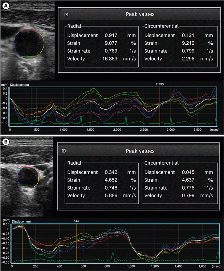
Figure 1 Strain analysis of the common carotid artery using the speckle tracking method for carotid B-mode ultrasonography in a 69-year-old normal control patient (A) and an age-matched ESRD patient (B). The strain and strain rate values were all decreased in the end-stage renal disease patient compared to the normal control patient.
Reference
-
1. Lindner A, Charra B, Sherrard DJ, Scribner BH. Accelerated atherosclerosis in prolonged maintenance hemodialysis. N Engl J Med. 1974; 290:697–701.
Article2. Briet M, Boutouyrie P, Laurent S, London GM. Arterial stiffness and pulse pressure in CKD and ESRD. Kidney Int. 2012; 82:388–400.
Article3. Georgianos PI, Sarafidis PA, Lasaridis AN. Arterial stiffness: a novel cardiovascular risk factor in kidney disease patients. Curr Vasc Pharmacol. 2015; 13:229–238.
Article4. London GM, Guérin AP, Marchais SJ, Métivier F, Pannier B, Adda H. Arterial media calcification in end-stage renal disease: impact on all-cause and cardiovascular mortality. Nephrol Dial Transplant. 2003; 18:1731–1740.
Article5. Kim SA, Park SM, Kim MN, et al. The relationship between mechanical properties of carotid artery and coronary artery disease. Eur Heart J Cardiovasc Imaging. 2012; 13:568–573.
Article6. Bjällmark A, Lind B, Peolsson M, Shahgaldi K, Brodin LA, Nowak J. Ultrasonographic strain imaging is superior to conventional non-invasive measures of vascular stiffness in the detection of age-dependent differences in the mechanical properties of the common carotid artery. Eur J Echocardiogr. 2010; 11:630–636.7. Yang EY, Brunner G, Dokainish H, et al. Application of speckle-tracking in the evaluation of carotid artery function in subjects with hypertension and diabetes. J Am Soc Echocardiogr. 2013; 26:901–9.e1.
Article8. Kidney Disease: Improving Global Outcomes (KDIGO) CKD-MBD Work Group. KDIGO clinical practice guideline for the diagnosis, evaluation, prevention, and treatment of Chronic Kidney Disease-Mineral and Bone Disorder (CKD-MBD). Kidney Int Suppl. 2009; S1–S130.9. Veronesi F, Corsi C, Caiani EG, Sarti A, Lamberti C. Tracking of left ventricular long axis from real-time three-dimensional echocardiography using optical flow techniques. IEEE Trans Inf Technol Biomed. 2006; 10:174–181.
Article10. Gamble G, Zorn J, Sanders G, MacMahon S, Sharpe N. Estimation of arterial stiffness, compliance, and distensibility from M-mode ultrasound measurements of the common carotid artery. Stroke. 1994; 25:11–16.
Article11. Zoungas S, Cameron JD, Kerr PG, et al. Association of carotid intima-medial thickness and indices of arterial stiffness with cardiovascular disease outcomes in CKD. Am J Kidney Dis. 2007; 50:622–630.
Article12. London GM, Guerin AP, Marchais SJ, et al. Cardiac and arterial interactions in end-stage renal disease. Kidney Int. 1996; 50:600–608.
Article13. Sigrist MK, Taal MW, Bungay P, McIntyre CW. Progressive vascular calcification over 2 years is associated with arterial stiffening and increased mortality in patients with stages 4 and 5 chronic kidney disease. Clin J Am Soc Nephrol. 2007; 2:1241–1248.
Article14. Raggi P, Bellasi A, Ferramosca E, Islam T, Muntner P, Block GA. Association of pulse wave velocity with vascular and valvular calcification in hemodialysis patients. Kidney Int. 2007; 71:802–807.
Article15. Blacher J, Guerin AP, Pannier B, Marchais SJ, Safar ME, London GM. Impact of aortic stiffness on survival in end-stage renal disease. Circulation. 1999; 99:2434–2439.
Article16. Yoon JH, Han D, Kim S, et al. Assessment of multidirectional movements of the common carotid artery in atherothrombotic stroke using dimensional speckle tracking carotid ultrasonography: A prospective, controlled cohort study. Echocardiography. 2018; 35:957–964.
Article17. Palmer SC, Hayen A, Macaskill P, et al. Serum levels of phosphorus, parathyroid hormone, and calcium and risks of death and cardiovascular disease in individuals with chronic kidney disease: a systematic review and meta-analysis. JAMA. 2011; 305:1119–1127.18. Briet M, Collin C, Karras A, et al. Arterial remodeling associates with CKD progression. J Am Soc Nephrol. 2011; 22:967–974.
Article19. Go AS, Chertow GM, Fan D, McCulloch CE, Hsu CY. Chronic kidney disease and the risks of death, cardiovascular events, and hospitalization. N Engl J Med. 2004; 351:1296–1305.
Article
- Full Text Links
-
- Actions
-
Cited
- CITED
-
- Close
- Share
-
- Similar articles
-
- Wall shear stress in hypertensive patients is associated with carotid vascular deformation assessed by speckle tracking strain imaging
- The Value of Elastic Modulus Index as a Novel Surrogate Marker for Cardiovascular Risk Stratification by Dimensional Speckle-Tracking Carotid Ultrasonography
- Non-invasive assessment of vascular alteration using ultrasound
- Speckle Tracking of Common Carotid Artery: A New Method for the Evaluation of Mechanical Vascular Function of Atherosclerosis
- Carotid ultrasound in patients with coronary artery disease
