Comparative Study on Acellular Dermal Graft Versus Propylene Mesh Both Either Loaded or Unloaded with BM-MSCs in Healing of Skull Bone Defect in Rats: Histological and Immunohistochemical Study
- Affiliations
-
- 1Department of Histology and Cell Biology, Faculty of Medicine Ain Shams University, Cairo, Egypt. ghada.hamam@yahoo.com
- 2Medical Research Center, Ain Shams University, Cairo, Egypt.
- KMID: 2429554
- DOI: http://doi.org/10.15283/ijsc18019
Abstract
- Bone defect occurs as a consequence of many conditions. Diseased bones don't heal properly and defects in face area need proper bone reconstruction to avoid psychological and social problems. Tissue engineering is an emerging new modality of treatment. We thought to study different methods to fill skull bone defect in rats in order to find the most safe and effective method. So, this study was designed to evaluate the efficacy of acellular dermal graft (ADM) versus propylene mesh both either loaded or unloaded with bone marrow derived mesenchymal stem cells (BM-MSCs) in healing of skull bone defect of a 5 mm diameter. The study included 36 adult male Wistar albino rats that were divided into three groups according to the way of filling skull bone defect. Group I: Ia (sham control), Ib (negative control). Group II: IIa (unseeded propylene), IIb (seeded propylene) and Group III: IIIa (unseeded ADM), IIIb (seeded ADM). The trephine operation was done on the left parietal bone. Specimens were collected four weeks postoperative and processed for H&E, osteopontin immunohistochemistry and scanning electron microscope. Morphometric and statistical analysis were also performed. After studying the results of the experiment, we found that propylene mesh and ADM were suitable scaffolds that could support new bone formation in clavarial bone defect. Healing of skull bone defect was better in rats that received seeded scaffolds more than rats with unseeded scaffolds. The seeded ADM showed significant increase in bone forming activity as confirmed by histomorphometric and statistical results.
Keyword
MeSH Terms
Figure
-

Fig. 1 (A) The defect after trephine. (B) ADM in the defect. (C~E) Phase contrast photomicrographs of MSCs showing (C) Sub-cultured MSCs on day 5. Most cells are spindle in shape with long cytoplasmic processes having granular cytoplasm with vesicular nuclei. (D&E) Cells of primary culture showing MSCs with positive brownish cytoplasmic immune reaction for CD 105 (↑) and CD44 respectively. (F) Propyelene mesh fibers (↑) in culture media with BM-MSCs (*). (G) H&E of ADM after seeding with BM-MSCs. Irregular collagen fibers in papillary (P) and reticular (R) layers are covered with basement membrane (↑). Inset: Cells (▲) between collagen fibers. ADM: acellular dermal graft, BM-MSCs: bone marrow derived mesenchymal stem cells. Phase contrast photomicrographs [C×200], [D×100], [E×400]. H&E [G×400, inset ×1,000].

Fig. 2 (A) Ia: Clavarial bone formed of diploe of compact bone with bone marrow space (BM) in-between. Endosteal surface lined with osteogenic cells (↑) and osteoblasts (↑↑), osteocytes inside their lacunae (O), and basophilic cement lines (▲) are seen between bone lamellae (*). (B~ D) Ib: (B) Bone defect filled with dense vascular (BV) connective tissue, few osteogenic cells (↑) at the margin of the defect, small foci of newly formed bone (↑↑). (C) Richly vascularized (BV) dense connective tissue in bone defect, fibroblasts (↑) between bundles of densely arranged collagen fibers (*). Occasional osteoblasts (▲) on bone surface (B). (D) A blood vessel is surrounded with homogenous acidophilic material (↑), mononuclear cells (▲), lymphocytes (thick arrow) and plasma cells (*). H&E [A, C&D] ×1,000. [B] ×400.
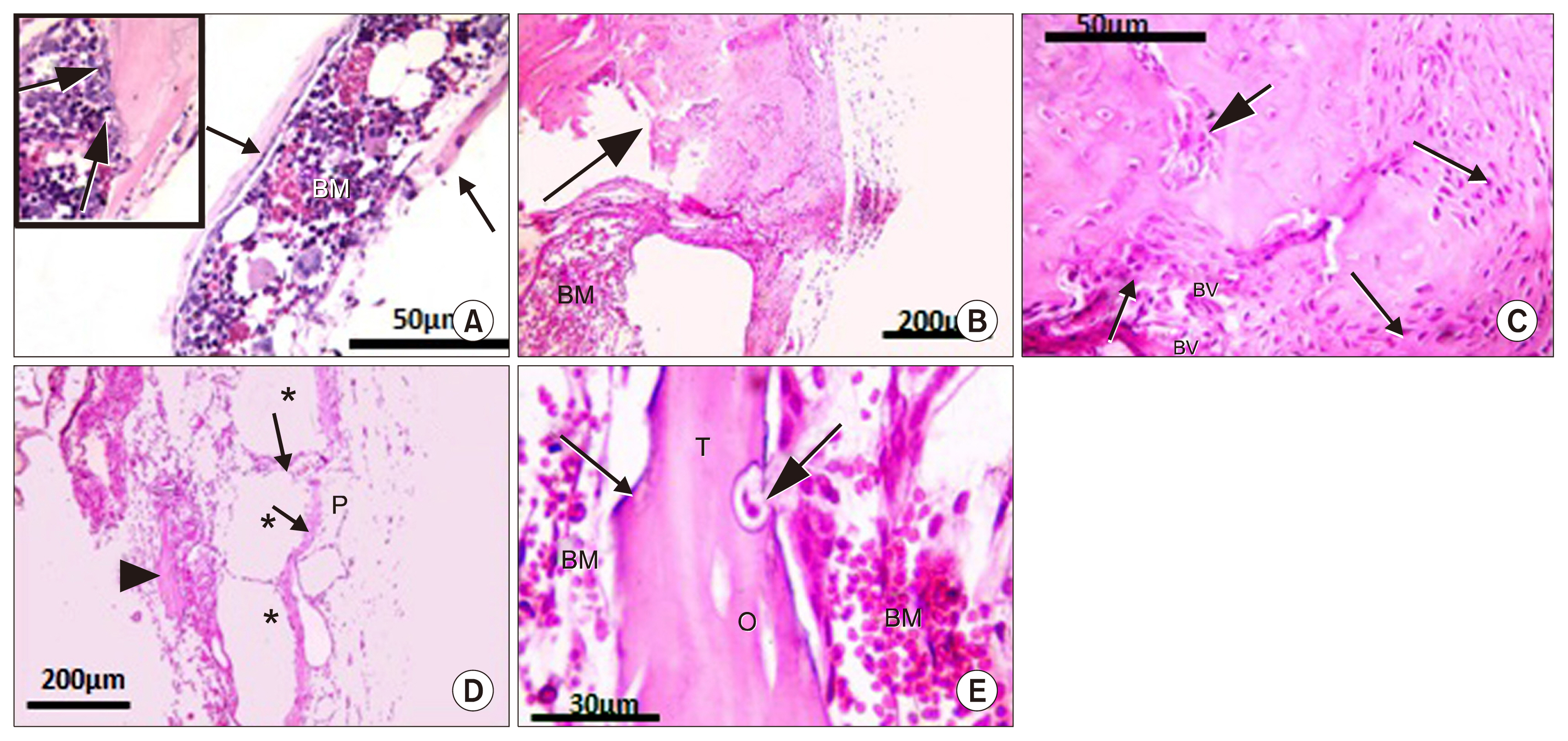
Fig. 3 (A) IIa: Thin rim of bone (↑) with attached cells on inner and outer surfaces of defect with bone marrow (BM) inbetween. Inset: margin of defect with osteogenic cells (thick arrow). (B~E) IIb: (B) Irregular immature bone (↑) and BM space near the defect. (C) Highly celluar bone margin, osteocytes in their lacunae, osteoblasts (↑), blood vessels (BV) and irrregulaly formed bone (thick arrow) near defect margin. (D) Bone defect filled with many wide spaces (*) separated by thin septa (↑) and covered with periosteum (P). Area of new bone formation is seen (▲). (E) Newly formed bone trabecula (T), oesteogenic cells (↑), osteocyte lacunae (O), an osteoclast (thick arrow) in its Hawship lacuna and BM cells. H&E [A, C&inset of A] ×400. [B&D] ×100. [E]×1,000.
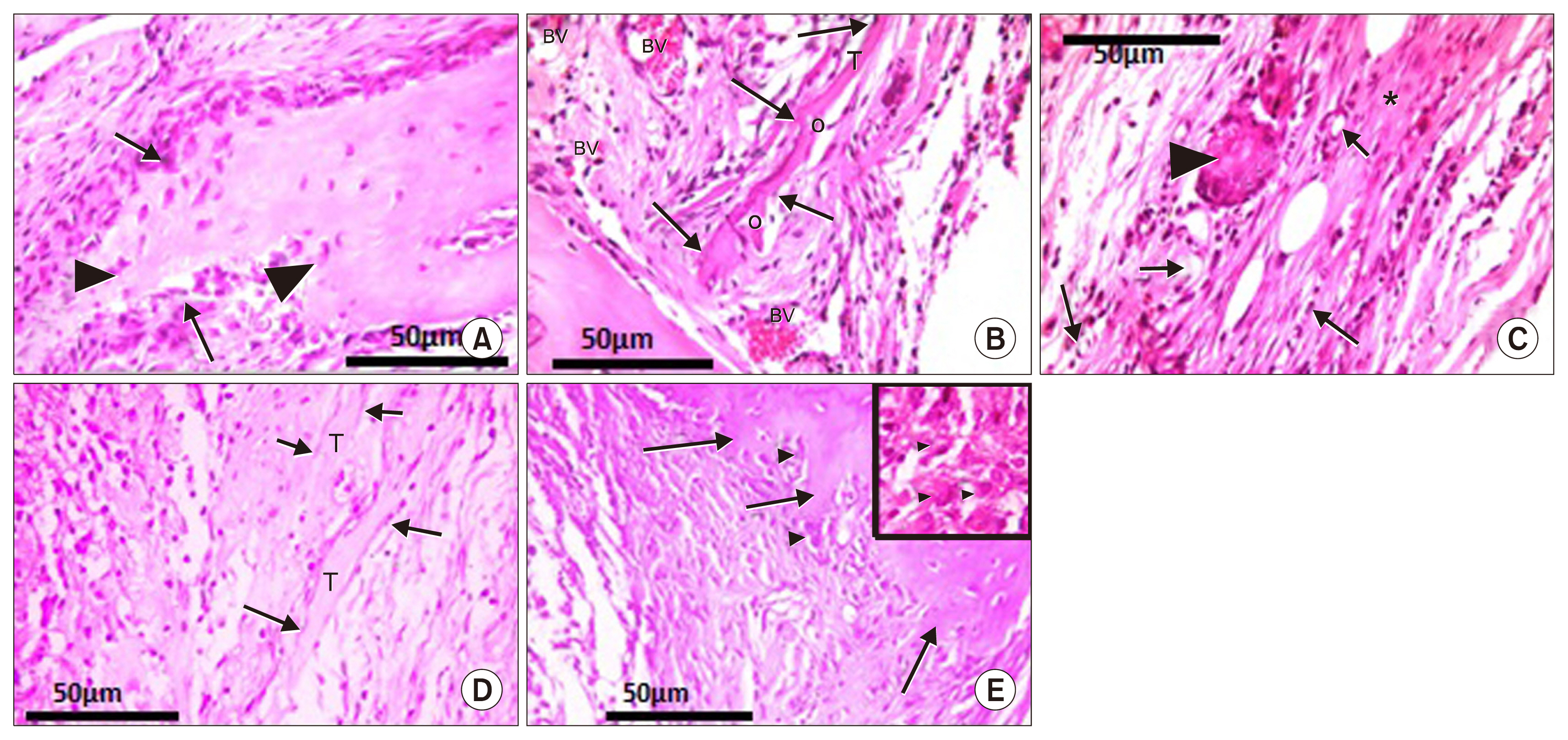
Fig. 4 (A~C) IIIa: (A) Highly cellular edge of defect, osteoblasts (↑), newly formed osteocytes (▲). (B) Highly vascular (BV) dermal graft, bone trabecula (T), osteocyte lacunae (O), osteogenic cells (↑). (C) Center of defect: dense connective tissue (*), newly formed bone (▲) surrounded with osteoblasts (c), osteogenic cells (f), blood vessels (↑). (D&E) IIIb: (D) Center of defect: Bone trabeculae (T) and osteogenic cells (↑). (E) Irregular margin of bone defect (↑) and osteoblasts (▲). Inset: Osteoblasts (▲) and highly cellular center of defect. H&E (A~E) ×400. [Inset of E] ×1,000.
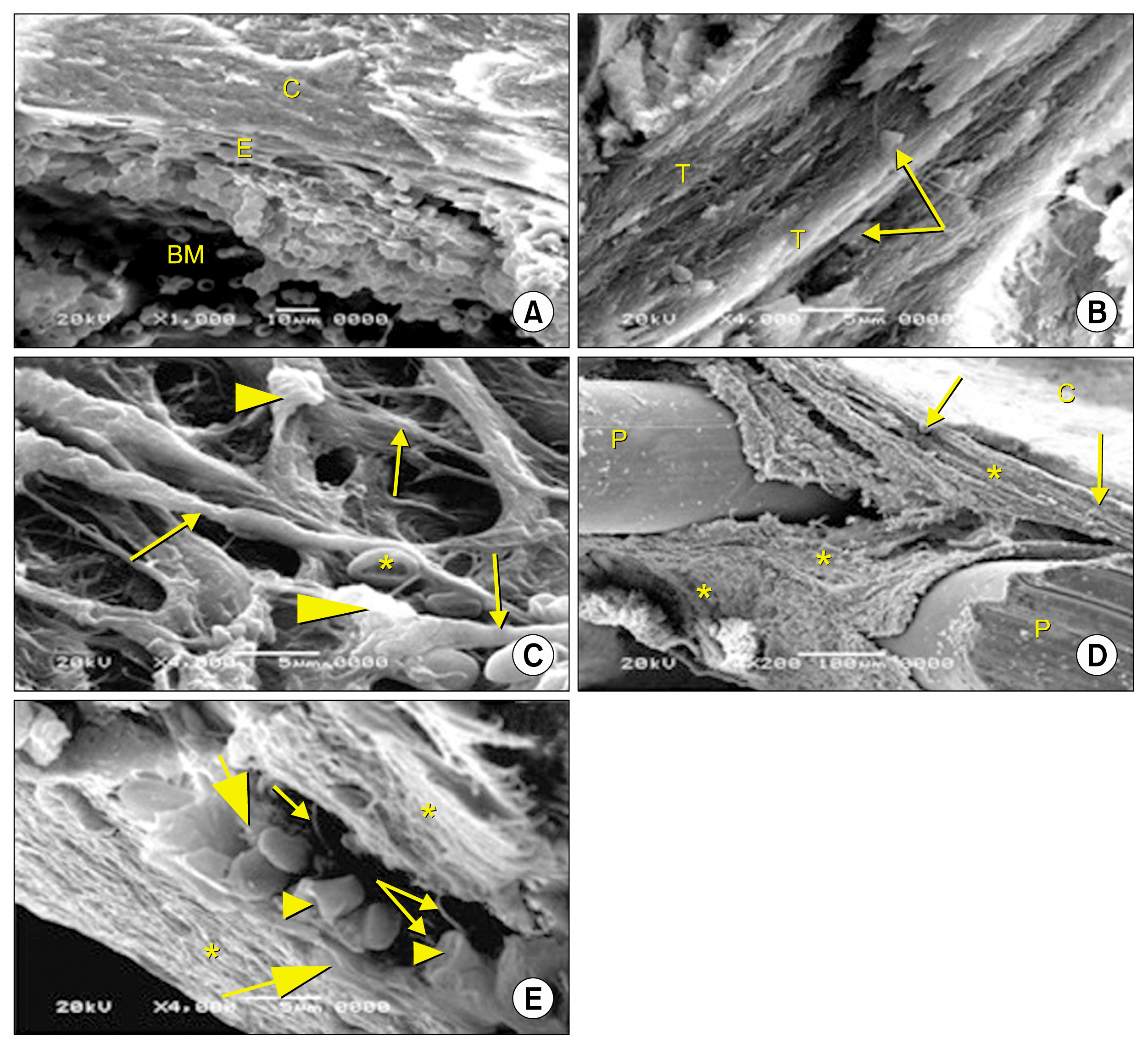
Fig. 5 Scanning electron micrographs of sagittal section of rat skull bone. (A&B) Ia: (A) Clavarial bone (C) enclosing bone marrow space (BM) is lined with flat endosteal cells (E). (B) Cuboidal bone cells (↑) on the surface of bone trabeculae (T). (C) Ib: Irregularly arranged collagen fibers (↑). Cells (▲) with long processes are attached to collagen fibers and RBCs (*) are seen inbetween. (D&E) IIa: (D) Fibers (*) and cells (↑) filling areas between propylene fibers (P). (C) Calvarial bone. (E) Regularly arranged fibers (*). Flattened cells (thick arrow) and cuboidal cells (▲) with cytoplasmic processes (↑) are seen between these fibers. SEM [A] ×1,000. [B, C&E] ×4,000. [D] ×200.
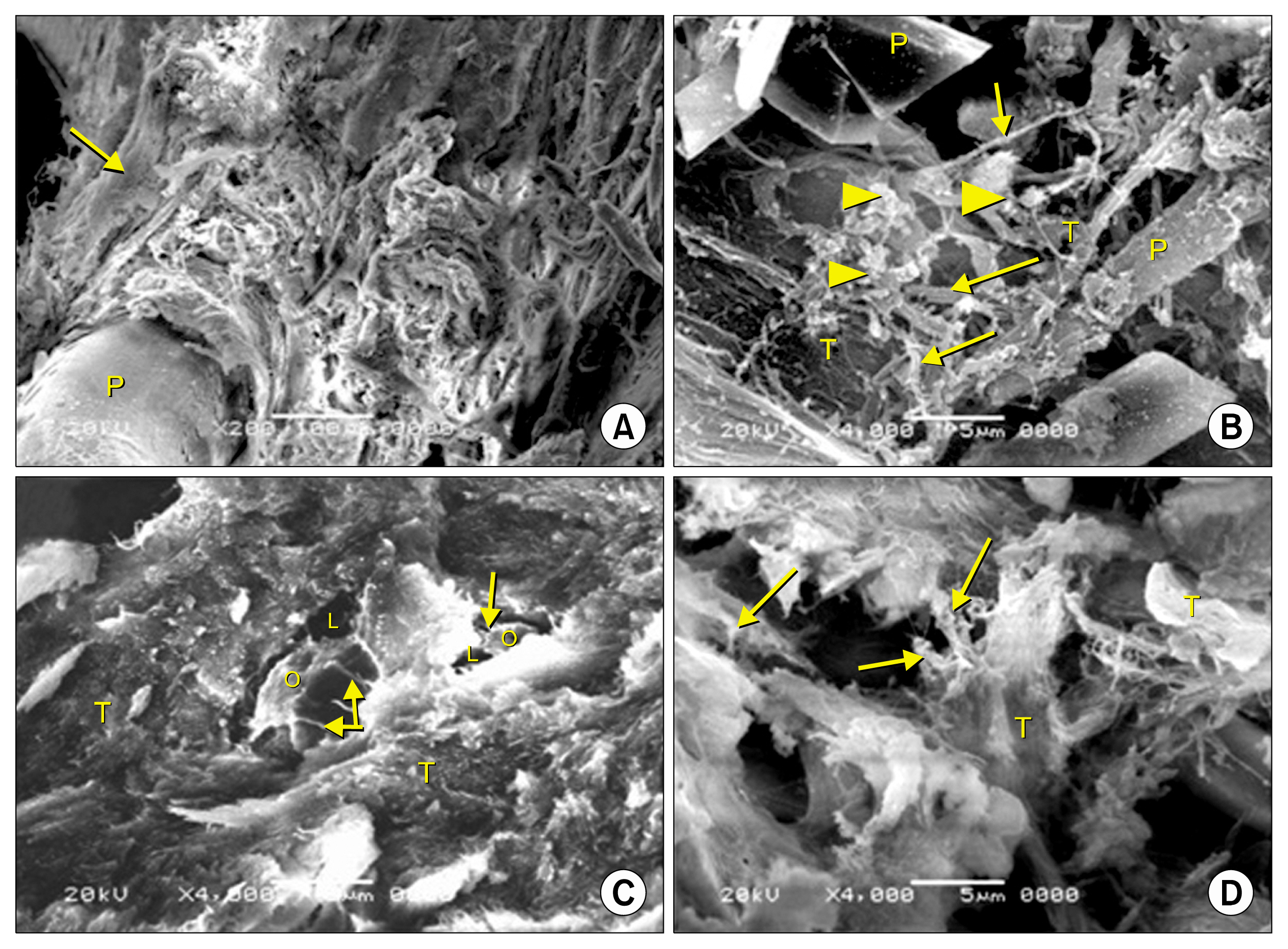
Fig. 6 Scanning electron micrographs of sagittal section of rat cranial vault at the defect area. (A&B) IIb: (A) A fiber of propylene mesh (P), surrounnded by irregular connective tissue filling the defect. Newly formed bone trabeculae can be seen at its periphery (↑). (B) Irregular fibers (↑), interconnected cells (▲) and irregular bone trabeculae (T) are seen between propylene fibers (P). (C) IIIa: Newly formed bone trabeculae (T) and osteocytes (O) inside their lacunae (L) can be seen with their processes (↑). (D) IIIb: The defect is filled with connective tissue. Irregular bone trabecula (T) and cells (↑) with interconnected processes can be seen between the trabeculae. SEM [A] ×200. [B, C&D] ×4,000.

Fig. 7 (A) Ia: Positive immune reaction at cement lines (↑), in cells lining the endosteum (▲) [from above downwards: osteogenic, osteoclast, osteoblasts. (B) Ib: Minimal expression in cells lining blood vessels (▲), at margin of defect (↑) and dura (D). (C) IIa: Positive reaction in cells at the margin of defect (↑) and on thin rim of bone on the inner and outer surfaces of defect (▲). (D&E) IIb: Positive immune reaction (D) in osteoblasts (▲) and osteocytes (▲), (E) In cells (↑) surrounding newly formed bone marrow space (BM). Inset: in cells invading the septa that separate bone marrow spaces. (F) IIIa: positive reaction in cells at defect margin (↑), in extracellular matrix (▲). Clavarial bone (B). (G&H) IIIb: Intense reaction in (G): extracellular matrix, osteoblasts (▲), osteocytes (▲), cements lines (↑), (H) in the center of the defect in cells (↑) and extracellular matrix (*). Anti osteopontin antibody [A~F&its inset, G&H] ×400.
Reference
-
References
1. Lewandrowski KU, Gresser JD, Wise DL, Trantol DJ. Bioresorbable bone graft substitutes of different osteoconductivities: a histologic evaluation of osteointegration of poly(propylene glycol-co-fumaric acid)-based cement implants in rats. Biomaterials. 2000; 21:757–764. DOI: 10.1016/S0142-9612(99)00179-9. PMID: 10721744.
Article2. Javaid MA, Kaartinen MT. Mesenchymal stem cell-based bone tissue engineering. International Dental Journal of Students Research. 2013; 1:24–35.3. Giannoudis PV, Dinopoulos H, Tsiridis E. Bone substitutes: an update. Injury. 2005; 36(Suppl 3):S20–S27. DOI: 10.1016/j.injury.2005.07.029. PMID: 16188545.
Article4. Chatterjea A, Meijer G, van Blitterswijk C, de Boer J. Clinical application of human mesenchymal stromal cells for bone tissue engineering. Stem Cells Int. 2010; DOI: 10.4061/2010/215625. PMID: 21113294. PMCID: 2989379.
Article5. Arnold S, Valerie J. Implant materials. Thorne CH, editor. Grabb and Smith’s Plastic Surgery. 6th ed. Lippincott Williams & Wilkins, a Wolters Kluwer business;2007. p. 58–65.6. Kaigler D, Pagni G, Park CH, Braun TM, Holman LA, Yi E, Tarle SA, Bartel RL, Giannobile WV. Stem cell therapy for craniofacial bone regeneration: a randomized, controlled feasibility trial. Cell Transplant. 2013; 22:767–777. DOI: 10.3727/096368912X652968.
Article7. Psarras S, Mavroidis M, Sanoudou D, Davos CH, Xanthou G, Varela AE, Panoutsakopoulou V, Capetanaki Y. Regulation of adverse remodelling by osteopontin in a genetic heart failure model. Eur Heart J. 2012; 33:1954–1963. DOI: 10.1093/eurheartj/ehr119.
Article8. Ferreira GR, Cestari TM, Granjeiro JM, Taga R. Lack of repair of rat skull critical size defect treated with bovine morphometric protein bound to microgranular bioabsorbable hydroxyapatite. Braz Dent J. 2004; 15:175–180. DOI: 10.1590/S0103-64402004000300002.
Article9. Spicer PP, Kretlow JD, Young S, Jansen JA, Kasper FK, Mikos AG. Evaluation of bone regeneration using the rat critical size calvarial defect. Nat Protoc. 2012; 7:1918–1929. DOI: 10.1038/nprot.2012.113. PMID: 23018195. PMCID: 3513397.
Article10. Mokal NJ, Desai MF. Calvarial reconstruction using high-density porous polyethylene cranial hemispheres. Indian J Plast Surg. 2011; 44:422–431. DOI: 10.4103/0970-0358.90812.
Article11. Walter JB, Talbot IC. Walter and Israel general pathology. 7th ed. British Library;1996. p. 184–415.12. Xu Y, Zhang G, Chang Y, Qiu YX, Wang C. The preparation of acellular dermal matrices by freeze-thawing and ultrasonication process and the evaluation of its antigenicity. Cell Biochem Biophys. 2015; 73:27–33. DOI: 10.1007/s12013-015-0569-9. PMID: 25649614.
Article13. McFarlin K, Gao X, Liu YB, Dulchavsky DS, Kwon D, Arbab AS, Bansal M, Li Y, Chopp M, Dulchavsky SA, Gautam SC. Bone marrow-derived mesenchymal stromal cells accelerate wound healing in the rat. Wound Repair Regen. 2006; 14:471–478. DOI: 10.1111/j.1743-6109.2006.00153.x. PMID: 16939576.
Article14. Li H, Fu X, Ouyang Y, Cai C, Wang J, Sun T. Adult bone-marrow-derived mesenchymal stem cells contribute to wound healing of skin appendages. Cell Tissue Res. 2006; 326:725–736. DOI: 10.1007/s00441-006-0270-9. PMID: 16906419.
Article15. Wang Q, Jin Y, Deng X, Liu H, Pang H, Shi P, Zhan Z. Second-harmonic generation microscopy for assessment of mesenchymal stem cell-seeded acellular dermal matrix in wound-healing. Biomaterials. 2015; 53:659–668. DOI: 10.1016/j.biomaterials.2015.03.011. PMID: 25890761.
Article16. Suvarna K, Layton C, Bancroft J. Theory and practice of histological techniques. 7th ed. USA: Churchill Livingston;2013.17. Armitage P, Berry G. Statistical methods in medical research. 3rd ed. Oxford: Blackwell Scientific Publications;1994. p. 12–48.18. Dorozhkin SV. Calcium orthophosphate-containing biocomposites and hybrid biomaterials for biomedical applications. J Funct Biomater. 2015; 6:708–832. DOI: 10.3390/jfb6030708. PMID: 26262645. PMCID: 4598679.
Article19. Ferreira LB, Bradaschia-Correa V, Moreira MM, Marques ND, Arana-Chavez VE. Evaluation of bone repair of critical size defects treated with simvastatin-loaded poly(lactic-coglycolic acid) microspheres in rat calvaria. J Biomater Appl. 2015; 29:965–976. DOI: 10.1177/0885328214550897.
Article20. Sohn JY, Park JC, Um YJ, Jung UW, Kim CS, Cho KS, Choi SH. Spontaneous healing capacity of rabbit cranial defects of various sizes. J Periodontal Implant Sci. 2010; 40:180–187. DOI: 10.5051/jpis.2010.40.4.180. PMID: 20827327. PMCID: 2931306.
Article21. Claes L, Recknagel S, Ignatius A. Fracture healing under healthy and inflammatory conditions. Nat Rev Rheumatol. 2012; 8:133–143. DOI: 10.1038/nrrheum.2012.1. PMID: 22293759.
Article22. Galindo P, Hernndez P, Padial M, Luisa M, Crespo V, O’Valle F. Osteopontin expression in an organic bovine bone Immunohistochemical osteopontin expression in bone xenograft in clinical series of maxillary sinus lift. J of Oral Sci & Rehab. 2015; 1:42–50.23. Cunniffe GM, Díaz-Payno PJ, Ramey JS, Mahon OR, Dunne A, Thompson EM, O’Brien FJ, Kelly DJ. Growth plate extracellular matrix-derived scaffolds for large bone defect healing. Eur Cell Mater. 2017; 33:130–142. DOI: 10.22203/eCM.v033a10. PMID: 28197989.
Article24. Zogbi L. The use of biomaterials to treat abdominal hernias. Biomaterials Applications for Nanomedicine. Pignatello Rosario, editor. 978-953-307-661-4. InTech;2011. p. 359–382.
Article25. Wolf K, Alexander S, Schacht V, Coussens LM, von Andrian UH, van Rheenen J, Deryugina E, Friedl P. Collagen-based cell migration models in vitro and in vivo. Semin Cell Dev Biol. 2009; 20:931–941. DOI: 10.1016/j.semcdb.2009.08.005. PMID: 19682592. PMCID: 4021709.
Article26. Petite H, Viateau V, Bensaïd W, Meunier A, de Pollak C, Bourguignon M, Oudina K, Sedel L, Guillemin G. Tissue-engineered bone regeneration. Nat Biotechnol. 2000; 18:959–963. DOI: 10.1038/79449. PMID: 10973216.
Article
- Full Text Links
-
- Actions
-
Cited
- CITED
-
- Close
- Share
-
- Similar articles
-
- Healing patterns of the acellular dermal matrix depend on graft method in the rabbit ears
- Usefulness of acellular dermal matrix graft on the tissue regeneration in rabbits
- Effects of Recombinant Human Bone Morphogenetic Protein-2 loaded Acellular Dermal Matrix on Bone Formation
- Autogenous Living Bone Graft and Dead Bone Graft in the Rabbit Tibia
- Role of Bone Marrow Mesenchymal Stem Cells in the Treatment of CCL4 Induced Liver Fibrosis in Albino Rats: A Histological and Immunohistochemical Study
