High-Pitch Coronary CT Angiography at 70 kVp Adopting a Protocol of Low Injection Speed and Low Volume of Contrast Medium
- Affiliations
-
- 1Department of Radiology, The First Affiliated Hospital of China Medical University, Shenyang 110001, P.R. China. 270973226@163.com
- 2Department of Radiology, Hebei General Hospital, Shijiazhuang 050051, P.R. China.
- KMID: 2427213
- DOI: http://doi.org/10.3348/kjr.2017.18.5.763
Abstract
OBJECTIVE
To evaluate the feasibility and image quality (IQ) of prospectively high-pitch coronary CT angiography (CCTA) with low contrast medium injection rate at 70 kVp.
MATERIALS AND METHODS
One hundred and four patients with suspected coronary artery disease (body mass index < 26 kg/m², sinus rhythm and heart rate < 70 beats/min) were prospectively enrolled and randomly divided into two groups. In group A and group B, 28 mL and 40 mL of 370 mgI/mL iodinated contrast media was administrated at a flow rate of 3.5 and 5 mL/s, respectively. CT values, noise, signal-to-noise ratio, contrast-to-noise ratio (CNR) of the proximal segments of coronary arteries and subjective IQ were evaluated.
RESULTS
The CT values and noise in group A were significantly lower than those in group B (434-485 Hounsfield units [HU] vs. 772-851 HU, all p < 0.001; 17.8-22.3 vs. 23.3-26.4, all p < 0.005). The CNRs of the right coronary artery and left main artery showed no statistical difference between the two groups (42.1 ± 13.8 vs. 36.8 ± 16.0, p = 0.074; 38.7 ± 10.6 vs. 38.1 ± 17.0, p = 0.819). No statistical difference was observed between the two groups in IQ scores (3.04 ± 0.75 vs. 3.0 ± 0.79, p = 0.526) and diagnostic ratio (96.1% [50/52] vs. 94.2% [49/52], p = 0.647).
CONCLUSION
Prospective high-pitch CCTA at 70 kVp with 28 mL of contrast media and injection rate of 3.5 mL/s could provide diagnostic IQ for normal-weight patients with heart rate of < 70 beats/min.
Keyword
MeSH Terms
Figure
-

Fig. 1 Flow chart for patient enrollment. CCTA = coronary CT angiography
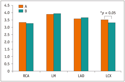
Fig. 2 Subjective ratings of CCTA IQ on coronary artery basis. LCX showed significant difference in IQ between two groups (p = 0.040). y-axis represent IQ score. A = group A, B = group B, IQ = image quality, LAD = left anterior descending artery, LCX = left circumflex artery, LM = left main coronary artery, RCA = right coronary artery
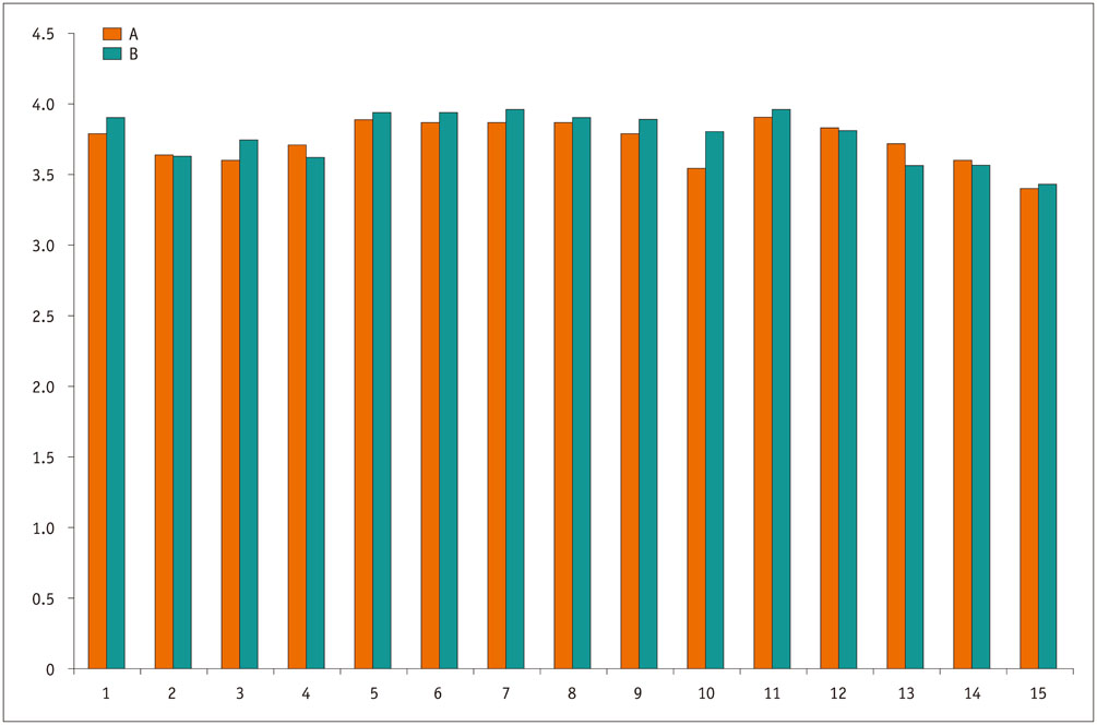
Fig. 3 Subjective ratings of CCTA IQ on coronary artery segment basis. x-axis represents coronary artery segments. y-axis represents IQ score. A = group A, B = group B.
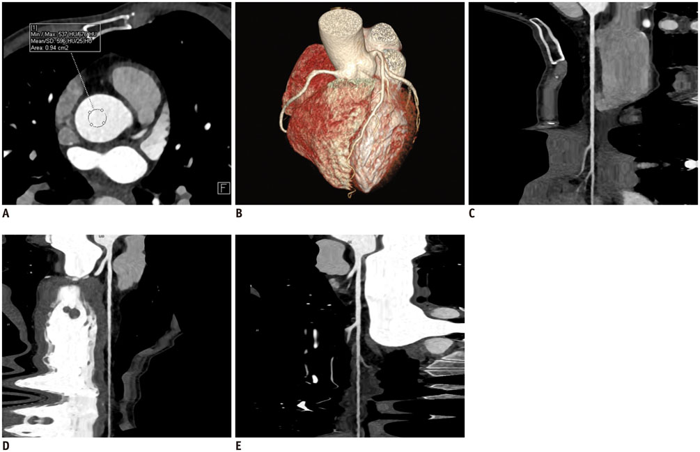
Fig. 4 65-year-old woman (HR = 56 bpm, BMI = 19.9 kg/m2, ED = 0.19 mSv) with atypical chest pain underwent CCTA. All visualized coronary segments were evaluated as score 4 (excellent IQ). A. CT value of AAO was 596 HU. B. Volume-rendered reformatted image of coronary tree. C-E. Curved multiplanar reformatted images of RCA, LAD, and LCX. AAO = ascending aorta root, BMI = body mass index, bpm = beats per minute, ED = effective dose, HR = heart rate, HU = Hounsfield unit

Fig. 5 CCTA in 74-year-old man (HR = 63 bpm, BMI = 24.3 kg/m2, ED = 0.38 mSv) with chest pain for one day. A. Showed CT value of AAO was 442 HU. B. Volume-rendered. C. Curved multiplanar reformation of RCA. D. Curved multiplanar reformation showed severe stenosis in proximal segment of LAD. E. Curved multiplanar reformation of LCX showed punctuate calcification in proximal segment. IQ of CCTA study was rated as score 4 (excellent) in this patient.
Cited by 1 articles
-
Age of Data in Contemporary Research Articles Published in Representative General Radiology Journals
Ji Hun Kang, Dong Hwan Kim, Seong Ho Park, Jung Hwan Baek
Korean J Radiol. 2018;19(6):1172-1178. doi: 10.3348/kjr.2018.19.6.1172.
Reference
-
1. Neefjes LA, Rossi A, Genders TS, Nieman K, Papadopoulou SL, Dharampal AS, et al. Diagnostic accuracy of 128-slice dual-source CT coronary angiography: a randomized comparison of different acquisition protocols. Eur Radiol. 2013; 23:614–622.2. Moscariello A, Vliegenthart R, Schoepf UJ, Nance JW Jr, Zwerner PL, Meyer M, et al. Coronary CT angiography versus conventional cardiac angiography for therapeutic decision making in patients with high likelihood of coronary artery disease. Radiology. 2012; 265:385–392.3. Litt HI, Gatsonis C, Snyder B, Singh H, Miller CD, Entrikin DW, et al. CT angiography for safe discharge of patients with possible acute coronary syndromes. N Engl J Med. 2012; 366:1393–1403.4. Kim YJ, Yong HS, Kim SM, Kim JA, Yang DH, Hong YJ, et al. Korean guidelines for the appropriate use of cardiac CT. Korean J Radiol. 2015; 16:251–285.5. von Ballmoos MW, Haring B, Juillerat P, Alkadhi H. Meta-analysis: diagnostic performance of low-radiation-dose coronary computed tomography angiography. Ann Intern Med. 2011; 154:413–420.6. Mitchell AM, Jones AE, Tumlin JA, Kline JA. Incidence of contrast-induced nephropathy after contrast-enhanced computed tomography in the outpatient setting. Clin J Am Soc Nephrol. 2010; 5:4–9.7. Morsbach F, Desbiolles L, Plass A, Leschka S, Schmidt B, Falk V, et al. Stenosis quantification in coronary CT angiography: impact of an integrated circuit detector with iterative reconstruction. Invest Radiol. 2013; 48:32–40.8. Komatsu S, Kamata T, Imai A, Ohara T, Takewa M, Ohe R, et al. Coronary computed tomography angiography using ultra-low-dose contrast media: radiation dose and image quality. Int J Cardiovasc Imaging. 2013; 29:1335–1340.9. Zhang C, Zhang Z, Yan Z, Xu L, Yu W, Wang R. 320-row CT coronary angiography: effect of 100-kV tube voltages on image quality, contrast volume, and radiation dose. Int J Cardiovasc Imaging. 2011; 27:1059–1068.10. Lu C, Wang Z, Ji J, Wang H, Hu X, Chen C. Evaluation of a chest circumference-adapted protocol for low-dose 128-slice coronary CT angiography with prospective electrocardiogram triggering. Korean J Radiol. 2015; 16:13–20.11. Achenbach S, Marwan M, Schepis T, Pflederer T, Bruder H, Allmendinger T, et al. High-pitch spiral acquisition: a new scan mode for coronary CT angiography. J Cardiovasc Comput Tomogr. 2009; 3:117–121.12. Lell MM, May M, Deak P, Alibek S, Kuefner M, Kuettner A, et al. High-pitch spiral computed tomography: effect on image quality and radiation dose in pediatric chest computed tomography. Invest Radiol. 2011; 46:116–123.13. Hou Y, Zheng J, Wang Y, Yu M, Vembar M, Guo Q. Optimizing radiation dose levels in prospectively electrocardiogram-triggered coronary computed tomography angiography using iterative reconstruction techniques: a phantom and patient study. PLoS One. 2013; 8:e56295.14. Zhang LJ, Qi L, Wang J, Tang CX, Zhou CS, Ji XM, et al. Feasibility of prospectively ECG-triggered high-pitch coronary CT angiography with 30 mL iodinated contrast agent at 70 kVp: initial experience. Eur Radiol. 2014; 24:1537–1546.15. Yeh BM, Shepherd JA, Wang ZJ, Teh HS, Hartman RP, Prevrhal S. Dual-energy and low-kVp CT in the abdomen. AJR Am J Roentgenol. 2009; 193:47–54.16. Mihl C, Kok M, Altintas S, Kietselaer BL, Turek J, Wildberger JE, et al. Evaluation of individually body weight adapted contrast media injection in coronary CT-angiography. Eur J Radiol. 2016; 85:830–836.17. Hell MM, Bittner D, Schuhbaeck A, Muschiol G, Brand M, Lell M, et al. Prospectively ECG-triggered high-pitch coronary angiography with third-generation dual-source CT at 70 kVp tube voltage: feasibility, image quality, radiation dose, and effect of iterative reconstruction. J Cardiovasc Comput Tomogr. 2014; 8:418–425.18. Den Harder AM, Willemink MJ, De Ruiter QM, De Jong PA, Schilham AM, Krestin GP, et al. Dose reduction with iterative reconstruction for coronary CT angiography: a systematic review and meta-analysis. Br J Radiol. 2016; 89:20150068.19. Cademartiri F, Maffei E, Arcadi T, Catalano O, Midiri M. CT coronary angiography at an ultra-low radiation dose (< 0.1 mSv): feasible and viable in times of constraint on healthcare costs. Eur Radiol. 2013; 23:607–613.20. Austen WG, Edwards JE, Frye RL, Gensini GG, Gott VL, Griffith LS, et al. A reporting system on patients evaluated for coronary artery disease. Report of the Ad Hoc Committee for Grading of Coronary Artery Disease, Council on Cardiovascular Surgery, American Heart Association. Circulation. 1975; 51:4 Suppl. 5–40.21. Chen MY, Shanbhag SM, Arai AE. Submillisievert median radiation dose for coronary angiography with a second-generation 320-detector row CT scanner in 107 consecutive patients. Radiology. 2013; 267:76–85.22. Bongartz G, Golding SJ, Jurik AG, Leonardi M, van Persijn E, van Meerten R, et al. European guidelines for multislice computed tomography: appendix c funded by the European Commission. Contract number FIGM-CT2000-20078-CT-TIP.Msct.eu Web site. Accessed October 12, 2015. http://www.msct.eu/CT_Quality_Criteria.htm.23. Halliburton SS, Abbara S, Chen MY, Gentry R, Mahesh M, Raff GL, et al. SCCT guidelines on radiation dose and dose-optimization strategies in cardiovascular CT. J Cardiovasc Comput Tomogr. 2011; 5:198–224.24. Cao JX, Wang YM, Lu JG, Zhang Y, Wang P, Yang C. Radiation and contrast agent doses reductions by using 80-kV tube voltage in coronary computed tomographic angiography: a comparative study. Eur J Radiol. 2014; 83:309–314.25. Zhang LJ, Qi L, De Cecco CN, Zhou CS, Spearman JV, Schoepf UJ, et al. High-pitch coronary CT angiography at 70 kVp with low contrast medium volume: comparison of 80 and 100 kVp high-pitch protocols. Medicine (Baltimore). 2014; 93:e92.26. Gordic S, Husarik DB, Desbiolles L, Leschka S, Frauenfelder T, Alkadhi H. High-pitch coronary CT angiography with third generation dual-source CT: limits of heart rate. Int J Cardiovasc Imaging. 2014; 30:1173–1179.27. Kidoh M, Nakaura T, Nakamura S, Namimoto T, Nozaki T, Sakaino N, et al. Contrast material and radiation dose reduction strategy for triple-rule-out cardiac CT angiography: feasibility study of non-ECG-gated low kVp scan of the whole chest following coronary CT angiography. Acta Radiol. 2014; 55:1186–1196.28. McCollough CH, Primak AN, Braun N, Kofler J, Yu L, Christner J. Strategies for reducing radiation dose in CT. Radiol Clin North Am. 2009; 47:27–40.29. LaBounty TM, Leipsic J, Poulter R, Wood D, Johnson M, Srichai MB, et al. Coronary CT angiography of patients with a normal body mass index using 80 kVp versus 100 kVp: a prospective, multicenter, multivendor randomized trial. AJR Am J Roentgenol. 2011; 197:W860–W867.30. Kok M, Mihl C, Hendriks BM, Altintas S, Kietselaer BL, Wildberger JE, et al. Optimizing contrast media application in coronary CT angiography at lower tube voltage: evaluation in a circulation phantom and sixty patients. Eur J Radiol. 2016; 85:1068–1074.31. Lell MM, Jost G, Korporaal JG, Mahnken AH, Flohr TG, Uder M, et al. Optimizing contrast media injection protocols in state-of-the art computed tomographic angiography. Invest Radiol. 2015; 50:161–167.32. Park EA, Lee W, Kang DK, Kim SJ, Kim YJ, Kim Y, et al. Comparison of iohexol-380 and iohexol-350 for coronary CT angiography: a multicenter, randomized, double-blind phase 3 trial. Korean J Radiol. 2016; 17:330–338.33. Achenbach S, Marwan M, Ropers D, Schepis T, Pflederer T, Anders K, et al. Coronary computed tomography angiography with a consistent dose below 1 mSv using prospectively electrocardiogram-triggered high-pitch spiral acquisition. Eur Heart J. 2010; 31:340–346.34. Husmann L, Valenta I, Gaemperli O, Adda O, Treyer V, Wyss CA, et al. Feasibility of low-dose coronary CT angiography: first experience with prospective ECG-gating. Eur Heart J. 2008; 29:191–197.
- Full Text Links
-
- Actions
-
Cited
- CITED
-
- Close
- Share
-
- Similar articles
-
- Technical Aspect of Coronary CT Angiography: Imaging Tips and Safety Issues
- The Effect of Non-Ionic Contrast Media on Q-T Interval and ST-T Wave of ECG during Coronary Angiography
- Accurate Measurement of Agatston Score Using kVp-Independent Reconstruction Algorithm for Ultra-High-Pitch Sn150 kVp CT
- The Image Quality and Radiation Dose of 100-kVp versus 120-kVp ECG-Gated 16-Slice CT Coronary Angiography
- Low Iodine Dose is Related with Overestimation of Extracellular Volume Derived from Cardiac CT
