Korean J Radiol.
2009 Jun;10(3):235-243. 10.3348/kjr.2009.10.3.235.
The Image Quality and Radiation Dose of 100-kVp versus 120-kVp ECG-Gated 16-Slice CT Coronary Angiography
- Affiliations
-
- 1Department of Radiology and the Institute of Radiation Medicine, Seoul National University College of Medicine, Seoul, 110-744, Korea. leew@radiol.snu.ac.kr
- 2Clinical Research Institute, Seoul National University Hospital, Seoul 110-744, Korea. leew@radiol.snu.ac.kr
- 3Department of Radiology, Seoul National University Hospital Healthcare System, Yeoksam-dong, Gangnam-gu, Seoul 135-984, Korea.
- KMID: 1779450
- DOI: http://doi.org/10.3348/kjr.2009.10.3.235
Abstract
OBJECTIVE
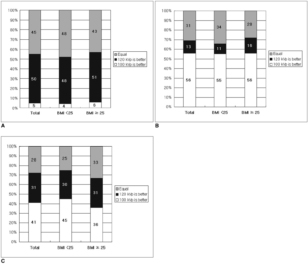
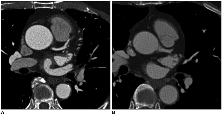
This study was conducted to assess the feasibility of performing 100-kVp electrocardiogram (ECG)-gated coronary CT angiography, as compared to 120-kVp ECG-gated coronary CT angiography. MATERIALS AND METHODS: We retrospectively evaluated one hundred eighty five gender- and body mass index-matched 16-slice coronary CT sets of data, which were obtained using either 100 kVp and 620 effective mAs or 120 kVp and 500 effective mAs. The density measurements (image noise, vessel density, signal-to-noise ratio [SNR] and contrast-to-noise ratio [CNR]) and the estimated radiation dose were calculated. As a preference test, two image readers were independently asked to choose one image from each pair of images. The results of both protocols were compared using the paired t-test or the Wilcoxon signed rank test. RESULTS: The 100-kVp images showed significantly more noise and a significantly higher vessel density than did the 120-kVp images. There were no significant differences in the SNR and CNR. The estimated reduction of the radiation dose for the 100-kVp protocol was 24%; 7.8 +/- 0.4 mSV for 100-kVp and 10.1 +/- 1.0 mSV for 120-kVp (p < 0.001). The readers preferred the 100-kVp images for reading (reader 1, p = 0.01; reader 2, p = 0.06), with their preferences being stronger when the subject's body mass index was less than 25. CONCLUSION: Reducing the tube kilovoltage from 120 to 100 kVp allows a significant reduction of the radiation dose without a significant change in the SNR and the CNR.
Keyword
MeSH Terms
-
Adult
Aged
Aged, 80 and over
Contrast Media/administration & dosage
Coronary Angiography/*methods
Electrocardiography/*methods
Feasibility Studies
Female
Humans
Iohexol/administration & dosage/analogs & derivatives
Male
Middle Aged
Observer Variation
*Radiation Dosage
Radiographic Image Enhancement/methods
Retrospective Studies
Tomography, X-Ray Computed/*methods
Figure
Reference
-
1. Ghersin E, Litmanovich D, Dragu R, Rispler S, Lessick J, Ofer A, et al. 16-MDCT coronary angiography versus invasive coronary angiography in acute chest pain syndrome: a blinded prospective study. AJR Am J Roentgenol. 2006. 186:177–184.2. Haberl R, Tittus J, Böhme E, Czernik A, Richartz BM, Buck J, et al. Multislice spiral computed tomographic angiography of coronary arteries in patients with suspected coronary artery disease: an effective filter before catheter angiography? Am Heart J. 2005. 149:1112–1119.3. Abada HT, Larchez C, Daoud B, Sigal-Cinqualbre A, Paul JF. MDCT of the coronary arteries: feasibility of low-dose CT with ECG-pulsed tube current modulation to reduce radiation dose. AJR Am J Roentgenol. 2006. 186:S387–S390.4. Hausleiter J, Meyer T, Hadamitzky M, Huber E, Zankl M, Martinoff S, et al. Radiation dose estimates from cardiac multislice computed tomography in daily practice: impact of different scanning protocols on effective dose estimates. Circulation. 2006. 113:1305–1310.5. d'Agostino AG, Remy-Jardin M, Khalil C, Delannoy-Deken V, Flohr T, Duhamel A, et al. Low-dose ECG-gated 64-slices helical CT angiography of the chest: evaluation of image quality in 105 patients. Eur Radiol. 2006. 16:2137–2146.6. Hohl C, Mühlenbruch G, Wildberger JE, Leidecker C, Süss C, Schmidt T, et al. Estimation of radiation exposure in low-dose multislice computed tomography of the heart and comparison with a calculation program. Eur Radiol. 2006. 16:1841–1846.7. Paul JF, Abada HT. Strategies for reduction of radiation dose in cardiac multislice CT. Eur Radiol. 2007. 17:2028–2037.8. Jakobs TF, Becker CR, Ohnesorge B, Flohr T, Suess C, Schoepf UJ, et al. Multislice helical CT of the heart with retrospective ECG gating: reduction of radiation exposure by ECG-controlled tube current modulation. Eur Radiol. 2002. 12:1081–1086.9. Hsieh J, Londt J, Vass M, Li J, Tang X, Okerlund D. Step-and-shoot data acquisition and reconstruction for cardiac x-ray computed tomography. Med Phys. 2006. 33:4236–4248.10. McCollough CH, Primak AN, Saba O, Bruder H, Stierstorfer K, Raupach R, et al. Dose performance of a 64-channel dual-source CT scanner. Radiology. 2007. 243:775–784.11. Earls JP, Berman EL, Urban BA, Curry CA, Lane JL, Jennings RS, et al. Prospectively gated transverse coronary CT angiography versus retrospectively gated helical technique: improved image quality and reduced radiation dose. Radiology. 2008. 246:742–753.12. Johnson TR, Nikolaou K, Wintersperger BJ, Leber AW, von Ziegler F, Rist C, et al. Dual-source CT cardiac imaging: initial experience. Eur Radiol. 2006. 16:1409–1415.13. Kalender WA, Wolf H, Suess C, Gies M, Greess H, Bautz WA. Dose reduction in CT by on-line tube current control: principles and validation on phantoms and cadavers. Eur Radiol. 1999. 9:323–328.14. Schoenhagen P. Back to the future: coronary CT angiography using prospective ECG triggering. Eur Heart J. 2008. 29:153–154.15. Stolzmann P, Leschka S, Scheffel H, Krauss T, Desbiolles L, Plass A, et al. Dual-source CT in step-and-shoot mode: noninvasive coronary angiography with low radiation dose. Radiology. 2008. 249:71–80.16. Stolzmann P, Scheffel H, Schertler T, Frauenfelder T, Leschka S, Husmann L, et al. Radiation dose estimates in dual-source computed tomography coronary angiography. Eur Radiol. 2008. 18:592–599.17. Heyer CM, Mohr PS, Lemburg SP, Peters SA, Nicolas V. Image quality and radiation exposure at pulmonary CT angiography with 100- or 120-kVp protocol: prospective randomized study. Radiology. 2007. 245:577–583.18. Sigal-Cinqualbre AB, Hennequin R, Abada HT, Chen X, Paul JF. Low-kilovoltage multi-detector row chest CT in adults: feasibility and effect on image quality and iodine dose. Radiology. 2004. 231:169–174.19. Wintersperger B, Jakobs T, Herzog P, Schaller S, Nikolaou K, Suess C, et al. Aorto-iliac multidetector-row CT angiography with low kV settings: improved vessel enhancement and simultaneous reduction of radiation dose. Eur Radiol. 2005. 15:334–341.20. Schueller-Weidekamm C, Schaefer-Prokop CM, Weber M, Herold CJ, Prokop M. CT angiography of pulmonary arteries to detect pulmonary embolism: improvement of vascular enhancement with low kilovoltage settings. Radiology. 2006. 241:899–907.21. Jung B, Mahnken AH, Stargardt A, Simon J, Flohr TG, Schaller S, et al. Individually weight-adapted examination protocol in retrospectively ECG-gated MSCT of the heart. Eur Radiol. 2003. 13:2560–2566.22. Huda W, Scalzetti EM, Levin G. Technique factors and image quality as functions of patient weight at abdominal CT. Radiology. 2000. 217:430–435.23. Kalra MK, Maher MM, Toth TL, Hamberg LM, Blake MA, Shepard JA, et al. Strategies for CT radiation dose optimization. Radiology. 2004. 230:619–628.24. Nakayama Y, Awai K, Funama Y, Liu D, Nakaura T, Tamura Y, et al. Lower tube voltage reduces contrast material and radiation doses on 16-MDCT aortography. AJR Am J Roentgenol. 2006. 187:W490–W497.
- Full Text Links
- Actions
-
Cited
- CITED
-
- Close
- Share
- Similar articles
-
- 100 kVp Low-Tube Voltage Abdominal CT in Adults: Radiation Dose Reduction and Image Quality Comparison of 120 kVp Abdominal CT
- Comparison of Image Quality of Shoulder CT Arthrography Conducted Using 120 kVp and 140 kVp Protocols
- Image Quality and Radiation Exposure in Coronary CT Angiography According to Tube Voltage and Body Mass Index
- High-Definition Computed Tomography for Coronary Artery Stent Imaging: a Phantom Study
- Effects of Dual-Energy CT with Non-Linear Blending on Abdominal CT Angiography


