J Korean Med Assoc.
2018 Nov;61(11):649-654. 10.5124/jkma.2018.61.11.649.
Role of dermoscopy and biopsy in the diagnosis of skin cancer: it takes two to tango
- Affiliations
-
- 1Department of Dermatology, Chung-Ang University College of Medicine, Seoul, Korea. ksli0209@cau.ac.kr
- KMID: 2426474
- DOI: http://doi.org/10.5124/jkma.2018.61.11.649
Abstract
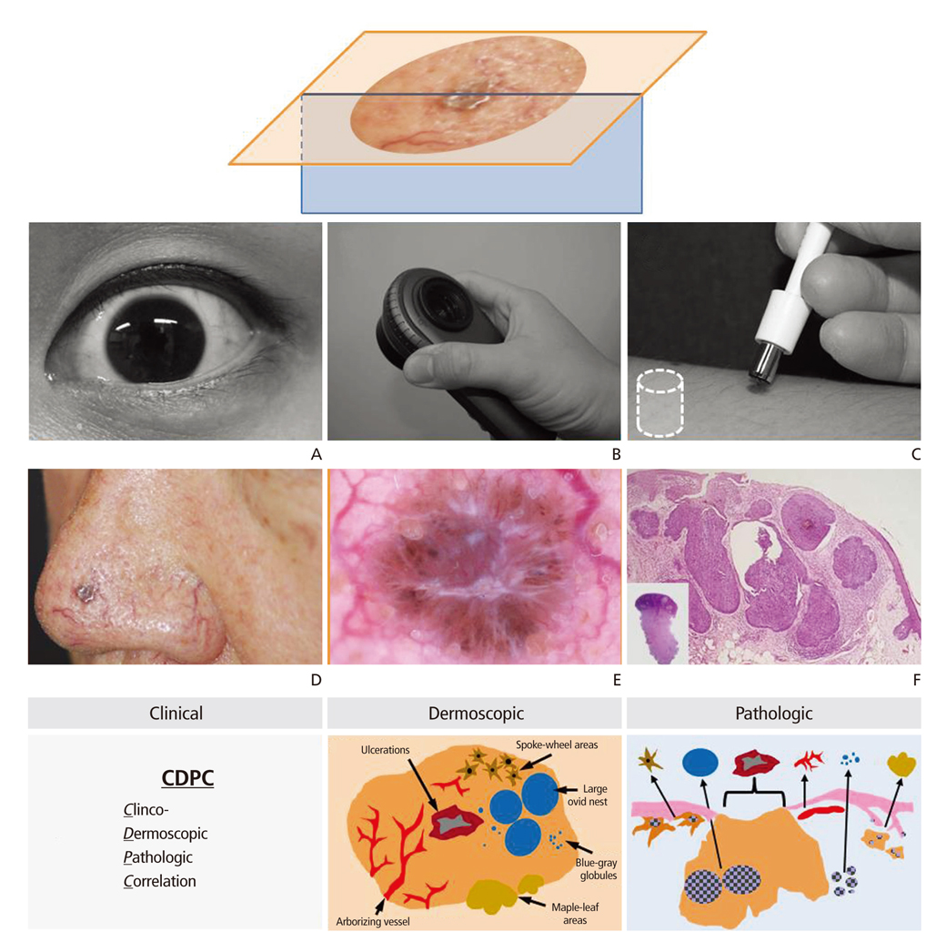
- Although the dermoscopy had very long history since its introduction in 17th century, only recently it has been possible to see the widespread application of dermoscopy in the dermatology clinic. One of the most promising areas where the dermoscopy can be applied is the diagnosis of skin cancer, especially malignant melanoma. Due to its inherent limitation to obtain in-depth information"”literally, from more than skin-deep and more importantly, from microscopic structures"”of skin cancers, dermoscopy cannot replace the present gold-standard "˜biopsy' in the diagnosis of skin cancer. However, several advantages of dermoscopy over biopsy merit further considerations. For example, as a non-invasive tool, dermoscopy is best suited for the follow-up of suspicious skin lesions, and as an all-at-a-glance tool, dermoscopy can aid the selection of the best biopsy-site to obtain the most meaningful pathological information from the minimal tissue specimen. There goes a saying that "˜it takes two to tango,' similarly, we might need the two (biopsy and dermoscopy) to cope rhythmically with the varying tempos of ever-progressing skin tumorigenesis and to reveal the true face of skin cancers usually hidden in various disguises.
Keyword
MeSH Terms
Figure
Reference
-
1. Hanahan D, Weinberg RA. Hallmarks of cancer: the next generation. Cell. 2011; 144:646–674.
Article2. Kapsokalyvas D, Bruscino N, Alfieri D, de Giorgi V, Cannarozzo G, Cicchi R, Massi D, Pimpinelli N, Pavone FS. Spectral morphological analysis of skin lesions with a polarization multispectral dermoscope. Opt Express. 2013; 21:4826–4840.
Article3. Yu C, Yang S, Kim W, Jung J, Chung KY, Lee SW, Oh B. Acral melanoma detection using a convolutional neural network for dermoscopy images. PLoS One. 2018; 13:e0193321.
Article4. Avci P, Gupta A, Sadasivam M, Vecchio D, Pam Z, Pam N, Hamblin MR. Low-level laser (light) therapy (LLLT) in skin: stimulating, healing, restoring. Semin Cutan Med Surg. 2013; 32:41–52.
- Full Text Links
- Actions
-
Cited
- CITED
-
- Close
- Share
- Similar articles
-
- The Diagnostic Accuracy of Dermoscopy for Scabies
- A Case of Superficial Basal Cell Carcinoma Not Correctly Diagnosed with Punch Biopsy but Diagnosed with Dermoscopy
- Xanthogranuloma for Whom Dermoscopy Was Used as an Adjuvant Diagnostic Tool
- Successfully Treated Basal Cell Carcinoma with Mohs Surgery, Diagnosing with Dermoscopy as a Primary Diagnostic Tool without Preoperative Punch Biopsy: A Report of Two Cases
- A Case of Amelanotic Melanoma: Dermoscopic Features


