Postpartum Superior Sagittal Sinus Thrombosis: A Case Report
- Affiliations
-
- 1Department of Neurosurgery, Hanyang University Guri Hospital, Guri, Korea. gksmh80@gmail.com
- KMID: 2424329
- DOI: http://doi.org/10.13004/kjnt.2018.14.2.146
Abstract
- Cerebral venous sinus thrombosis (CVST) is a rare disease. Early diagnosis and treatment are important, as CVST is potentially fatal. Pregnancy and puerperium are known risk factors for CVST. Here, we report the case of a patient who developed superior sagittal sinus thrombosis after a normal vaginal delivery. A 20-year-old woman presented with a headache and seizures two days after a normal vaginal delivery. Initially, brain computed tomography (CT) showed a subarachnoid hemorrhage in the right parietal lobe and sylvian fissure, together with mild cerebral edema. CT angiography revealed superior sagittal sinus thrombosis. Multiple micro-infarctions were seen on diffusion-weighted magnetic resonance images. An intravenous infusion of heparin and mannitol was administered immediately. Two days after treatment initiation, the patient showed sudden neurological deterioration, with left-sided hemiplegia. Brain CT showed moderate brain edema and hemorrhagic densities. Emergency decompressive craniectomy was performed, and heparin was re-administered on post-operative day (POD) 1. On POD 9, the patient's mental state improved from stupor to drowsy, but the left-sided hemiplegia persisted. CT angiography showed that the superior sinus thrombosis had decreased. Superior sagittal sinus thrombosis is an uncommon complication, with an unfavorable outcome, after delivery. Timely diagnosis and treatment are important for preventing neurological deterioration.
MeSH Terms
-
Angiography
Brain
Brain Edema
Cerebral Infarction
Decompressive Craniectomy
Diagnosis
Early Diagnosis
Emergencies
Female
Headache
Hemiplegia
Heparin
Humans
Infusions, Intravenous
Mannitol
Parietal Lobe
Postpartum Period*
Pregnancy
Rare Diseases
Risk Factors
Seizures
Sinus Thrombosis, Intracranial
Stupor
Subarachnoid Hemorrhage
Superior Sagittal Sinus*
Thrombosis*
Young Adult
Heparin
Mannitol
Figure
-
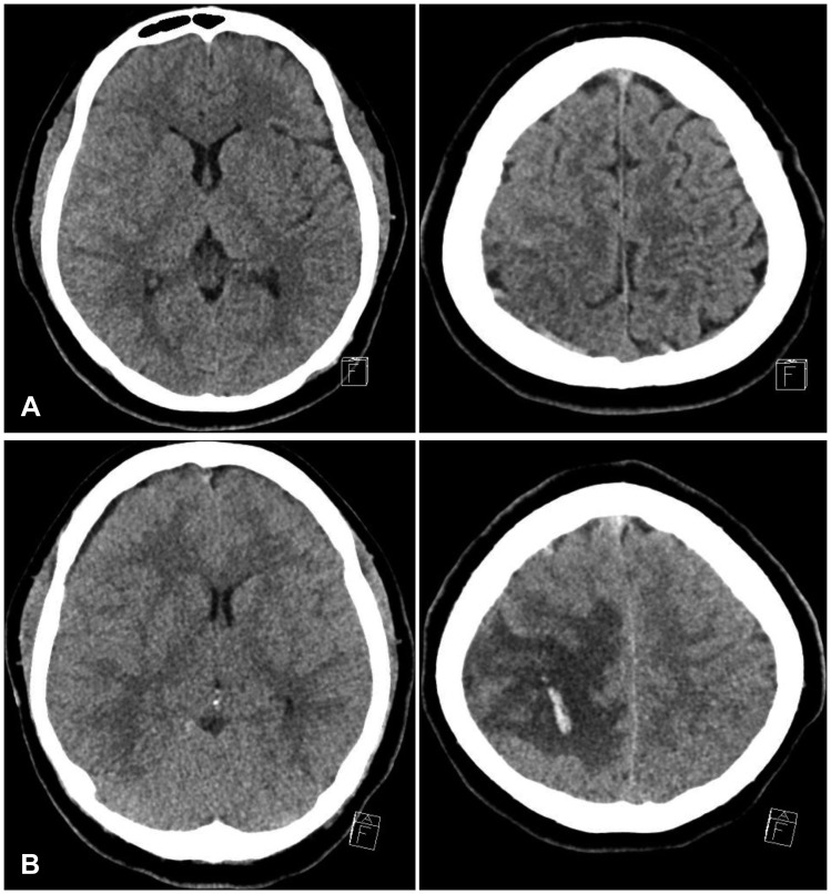
FIGURE 1 (A) Initial brain computer tomography (CT) shows acute infarct in the right high parietal lobe subcortical/deep white matter and left cerebellar hemisphere. Also evident is subarachnoid hemorrhage on the right sylvian fissure, and frontotemporal convexity sulci. There is no apparent midline shift before exacerbation. (B) Follow-up brain CT shows the aggravating venous infarct, with focal hemorrhage on the right high frontoparietal lobe, and swelling after exacerbation.
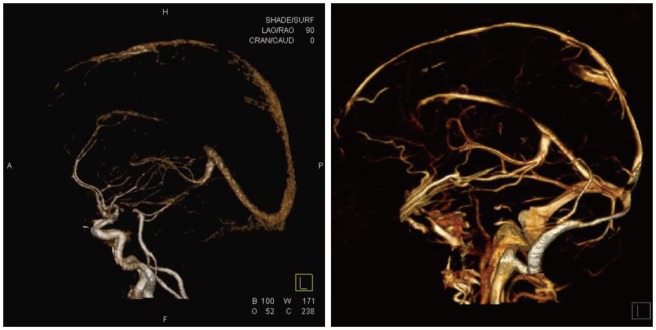
FIGURE 2 Computer tomography (CT)-digital subtraction angiography shows more resolving progression, interval decreased extent of superior sagittal sinus thrombosis (left: initial CT angiography, right: follow-up CT angiography).
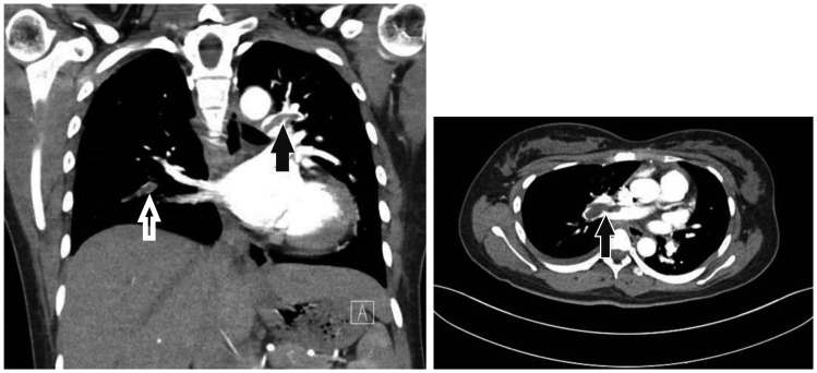
FIGURE 3 Chest computer tomography angiography shows pulmonary thromboembolism at the pulmonary trunk bifurcation, and both pulmonary arteries in the right lower, left upper and left lower lobes.
Reference
-
1. Borum SE, Naul LG, McLeskey CH. Postpartum dural venous sinus thrombosis after postdural puncture headache and epidural blood patch. Anesthesiology. 1997; 86:487–490. PMID: 9054267.
Article2. Ehtisham A, Stern BJ. Cerebral venous thrombosis: a review. Neurologist. 2006; 12:32–38. PMID: 16547444.3. Ferro JM, Canhão P, Stam J, Bousser MG, Barinagarrementeria F. Prognosis of cerebral vein and dural sinus thrombosis: results of the International Study on Cerebral Vein and Dural Sinus Thrombosis (ISCVT). Stroke. 2004; 35:664–670. PMID: 14976332.4. Ghandehari K, Riasi HR, Noureddine A, Masoudinezhad S, Yazdani S, Mirzae MM, et al. Safety assessment of anticoagulation therapy in patients with hemorrhagic cerebral venous thrombosis. Iran J Neurol. 2013; 12:87–91. PMID: 24250911.5. Gross PL, Weitz JI. New anticoagulants for treatment of venous thromboembolism. Arterioscler Thromb Vasc Biol. 2008; 28:380–386. PMID: 18296593.
Article6. Lanska DJ, Kryscio RJ. Risk factors for peripartum and postpartum stroke and intracranial venous thrombosis. Stroke. 2000; 31:1274–1282. PMID: 10835444.
Article
- Full Text Links
-
- Actions
-
Cited
- CITED
-
- Close
- Share
-
- Similar articles
-
- A Case of Eosinophilic Panniculitis Associated With Superior Sagittal Sinus Thrombosis
- Thrombosis of the Superior Sagittal Sinus in Behcet's Disease With Vascular and Enteric Involvements
- Transient Neurologic Deterioration after Total Removal of Parasagittal Meningioma Including Completely Occluding Superior Sagittal Sinus
- Cerebral Venous Sinus Thrombosis in an Adolescent Presenting with Headache
- Superior sagittal sinus dural arteriovenous fistula caused by treatment of meningioma masquerades as sinus thrombosis
