Metaplastic Meningioma Overspreading the Cerebral Convexity
- Affiliations
-
- 1Department of Neurosurgery, Ilsan Paik Hospital, College of Medicine, Inje University, Goyang, Korea. cychoi@paik.ac.kr
- 2Department of Pathology, Ilsan Paik Hospital, College of Medicine, Inje University, Goyang, Korea.
- KMID: 2423982
- DOI: http://doi.org/10.14791/btrt.2018.6.e17
Abstract
- Meningioma is relatively common, benign, and extra-axial tumor accounting for about 20% of primary brain and spinal cord tumors. The World Health Organization (WHO) classified these tumors into Grade I (benign), Grade II (atypical), and Grade III (anaplastic) meningioma. Grade I meningioma which is slowly growing tumor and have some rare subtypes. Among them, metaplastic subtype is defined as a tumor containing focal or widespread mesenchymal components including osseous, cartilaginous, lipomatous, myxoid or xanthomatous tissue, singly or in combinations. We report a rare metaplastic meningioma overspreading nearly whole cerebral convexity from main extra-axial tumor bulk in the parietal lobe.
MeSH Terms
Figure
-
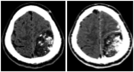
Fig. 1 Brain CT shows about 6.7×4.5 cm sized, low-density, broad dural based, and inhomogeneous enhancing mass with multiple calcifications in the left parietal region which is suggesting extra-axial tumor. Definite subdural lesion is not seen on the brain CT.
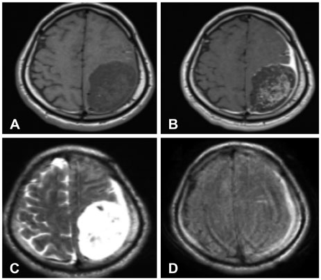
Fig. 2 Brain MRI shows extra-axial inhomogeneous enhancing tumor in the left fronto-parietal region. Bulky parietal mass shows low-signal intensity and the other flat subdural mass overspreading cerebral convexity shows iso-signal intensity on T1-weighted image (WI) (A). Inhomogeneous and homogeneous enhancements are seen in bulky parietal mass and flat subdural mass respectively (B). High-signal intensity in bulky parietal and flat subdural mass are seen on T2-WI (C). On fluid attenuated inversion recovery image, iso-signal intensity in the bulky parietal mass and high-signal intensity in the flat subdural mass are seen (D).
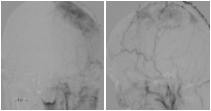
Fig. 3 On the cerebral angiograms, tumor staining via the left middle meningeal artery is seen.
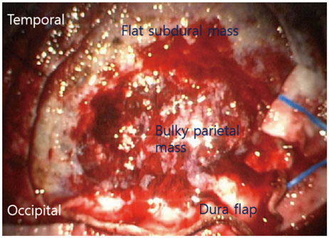
Fig. 4 Operative findings. Bulky and soft tumor mass with multiple small cysts and calcifications are seen in the parasagittal space of left parietal lobe. Flat and rubbery tumor mass in the subdural space is overspreading the nearly whole cerebral convexity. These tumors are totally extra-axial. And the tumors were not infiltrated to the brain parenchyme.
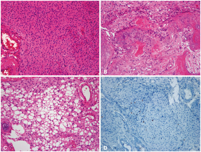
Fig. 5 Pathologic findings. A: The tumor composed of plump, elongated cells with meningothelial features in syncytial arrangement. The nuclei are round to ovoid harboring fine chromatin and inconspicuous nucleoli. There is no evidence of mitosis or necrosis (hematoxylin-eosin staining, ×10). B: The areas of chicken-wire-like calcification and ossification are seen in several areas (hematoxylin-eosin staining, ×40). C: Lipomatous metaplasia and xanthomatous change are present (hematoxylin-eosin staining, ×100). D: The tumor cells show a low Ki-67 labelling index with about 1% (immunohistochemistry staining, ×40).
Cited by 1 articles
-
Intracranial Metaplastic Meningioma : Clinical and Radiological Characteristics of 11 Cases
Taehoon Kim, Jin Wook Kim, So Young Ji, Ho Kang, Kyung-Min Kim, Yong Hwy Kim, Chul-Kee Park, Seung Hong Choi, Sung-Hye Park
J Korean Neurosurg Soc. 2020;63(5):657-663. doi: 10.3340/jkns.2020.0151.
Reference
-
1. Perry A, Louis DN, Scheithauer BW, Budka H, von Deimling A. Meningiomas. In : Louis DN, Ohgaki H, Wiestler OD, Cavenee WK, editors. World Health Organization classification of tumours of the central nervous system. Lyon: IARC Press;2007. p. 164–172.2. Huang J, Petersson F. Intracerebral metaplastic meningioma with prominent ossification and extensive calcification. Rare Tumors. 2011; 3:e20.
Article3. Tang H, Sun H, Chen H, et al. Clinicopathological analysis of metaplastic meningioma: report of 15 cases in Huashan Hospital. Chin J Cancer Res. 2013; 25:112–118.4. Matyja E, Naganska E, Zabek M, Jagielski J. Meningioma with the unique coexistence of secretory and lipomatous components: a case report with immunohistochemical and ultrastructural study. Clin Neuropathol. 2005; 24:257–261.5. Johnson MD, Stevenson CB, Thompson RC, Atkinson J, Boyer P. December 2006: 31-year-old woman with hemiparesis. Brain Pathol. 2007; 17:255–257.
Article6. Majumdar K, Mandal S, Thakkar R, Saran RK, Srivastava AK. Meningeal osteochondroma simulating meningioma with metaplastic change: a rare golf-ball-like lesion of non-meningothelial mesenchymal origin. Brain Tumor Pathol. 2014; 31:62–67.
Article
- Full Text Links
-
- Actions
-
Cited
- CITED
-
- Close
- Share
-
- Similar articles
-
- Intracranial Metaplastic Meningioma : Clinical and Radiological Characteristics of 11 Cases
- Cerebral Arteriovenous Malformation Associated with Intracranial Meningioma and Aneurysm: Case Report
- Convexity Meningioma En Plaque Presenting with Diffuse Hyperostosis of the Skull
- Anaplastic Cystic Meningioma
- Totally Ossified Metaplastic Spinal Meningioma
