Totally Ossified Metaplastic Spinal Meningioma
- Affiliations
-
- 1Department of Neurosurgery, Hokkaido University, Sapporo, Japan. jchangil@chosun.ac.kr
- 2Department of Neurosurgery, School of Medicine, Chosun University, Gwangju, Korea.
- KMID: 2190910
- DOI: http://doi.org/10.3340/jkns.2013.54.3.257
Abstract
- A 61-year-old woman with a very rare case of totally ossified large thoracic spinal metaplastic meningioma, showing progressing myelopathy is presented. Computed tomographic images showed a large totally ossfied intradural round mass occupying the spinal canal on T9-10 level. Magnetic resonance imaging revealed a large T9-10 intradural extramedullary mass that was hypointense to spinal cord on T1- and T2-weighted sequences, partial enhancement was apparent after Gadolinium administration. The spinal cord was severely compressed and displaced toward the right at the level of T9-10. Surgical removal of the tumor was successfully accomplished via the posterior midline approach and the histological diagnosis verified an ossified metaplastic meningioma. The clinical neurological symptoms of patient were improved postoperatively. In this article we discuss the surgical and pathological aspects of rare case of spinal totally ossified metaplastic meningioma.
MeSH Terms
Figure
-
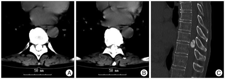
Fig. 1 Computed tomographic scan without enhancement show a large totally ossified intradural round mass occupying the whole spinal canal on T9-T10 level. Axial view on T9 (A),10 level (B) and sagittal view (C).

Fig. 2 Magnetic resonance image showing the tumor was very low signal intensity on T2-weighted image and mainly low intensity compared to spinal cord on T1-weighted image (A and B). T1-weighted imaging gadolinium showed a round homogenously enhanced tumor and the lower signal intensity of central tumor portions were less enhanced compared with the other portions of the masses (C).

Fig. 3 In the totally ossified metaplastic meningioma. It is difficult to find the arachnoid membrane that acts a natural barrier and provides a dissection plane during surgery (A). This tumor has been dissected meticulously through the adhesive peritumoral arachnoid membrane with microsurgical scissor and en-bloc technique (B).

Fig. 4 Histologic examination of the initially resected tumor specimen stained with hematoxylin and eosin shows metaplastic (osseous) meningioma. A : The tumor cells which have delicate oval nuclei, inconspicuous nucleoli, and lightly eosinophilic cytoplasm, proliferate forming cellular whorls (×40). B : Within the tumor, marked heterotopic ossification occurs without psammoma bodies (×10).
Reference
-
1. Antons K. Calcified spinal meningioma visible on the roentgen film. Acta Psychiat Scand. 1944; 19:5–9.
Article2. Bonstelle CT, Vines FS. Calcification in a cervical intraspinal neurilemmoma. Neuroradiology. 1976; 10:231–233. PMID: 934461.
Article3. Camp JD. The roentgenologic localization of tumors affecting the spinal cord. Am J Roentgenol. 1938; 40:540–544.4. Calogero JA, Moossy J. Extradural spinal meningiomas. Report of four cases. J Neurosurg. 1972; 37:442–447. PMID: 5070871.5. Culver GJ, Concannon JP, Koenig EC. Calcification in intraspinal meningiomas. Am J Roentgenol Radium Ther. 1949; 62:237–246.6. Doita M, Harada T, Nishida K, Marui T, Kurosaka M, Yoshiya S. Recurrent calcified spinal meningioma detected by plain radiograph. Spine (Phila Pa 1976). 2001; 26:E249–E252. PMID: 11389409.
Article7. Freidberg SR. Removal of an ossified ventral thoracic meningioma. Case report. J Neurosurg. 1972; 37:728–730. PMID: 4631926.8. Garfinkle W, Yudd AP. Calcified intraspinal meningioma detected by computed tomography. Comput Radiol. 1982; 6:305–307. PMID: 7172649.
Article9. Gezen F, Kahraman S, Canakci Z, Bedük A. Review of 36 cases of spinal cord meningioma. Spine (Phila Pa 1976). 2000; 25:727–731. PMID: 10752106.
Article10. Huang TY, Kochi M, Kuratsu J, Ushio Y. Intraspinal osteogenic meningioma : report of a case. J Formos Med Assoc. 1999; 98:218–221. PMID: 10365544.11. Kaufman AB, Dunsmore RH. Clinicopathological considerations in spinal meningeal calcification and ossification. Neurology. 1971; 21:1243–1248. PMID: 5002424.
Article12. King AT, Sharr MM, Gullan RW, Bartlett JR. Spinal meningiomas : a 20-year review. Br J Neurosurg. 1998; 12:521–526. PMID: 10070460.13. Kitagawa M, Nakamura T, Aida T, Iwasaki Y, Abe H, Nagashima K. [Clinicopathologic analysis of ossification in spinal meningioma]. Noshuyo Byori. 1994; 11:115–119. PMID: 8162148.14. Klekamp J, Samii M. Surgical results for spinal meningiomas. Surg Neurol. 1999; 52:552–562. PMID: 10660020.
Article15. Kubota T, Sato K, Yamamoto S, Hirano A. Ultrastructural study of the formation of psammoma bodies in fibroblastic meningioma. J Neurosurg. 1984; 60:512–517. PMID: 6699695.
Article16. Levy WJ Jr, Bay J, Dohn D. Spinal cord meningioma. J Neurosurg. 1982; 57:804–812. PMID: 7143063.
Article17. Memon MY, Schneck L. Ventral spinal tumor : the value of computed tomography in its localization. Neurosurgery. 1981; 8:108–111. PMID: 7207766.18. Mirimanoff RO, Dosoretz DE, Linggood RM, Ojemann RG, Martuza RL. Meningioma : analysis of recurrence and progression following neurosurgical resection. J Neurosurg. 1985; 62:18–24. PMID: 3964853.
Article19. Naderi S, Yilmaz M, Canda T, Acar U. Ossified thoracic spinal meningioma in childhood : a case report and review of the literature. Clin Neurol Neurosurg. 2001; 103:247–249. PMID: 11714573.20. Niijima K, Huang YP, Malis LI, Sachdev VP. Ossified spinal meningioma en plaque. Spine (Phila Pa 1976). 1993; 18:2340–2343. PMID: 8278859.
Article21. Nyström SH, Nyholm M. The origin of the calcium deposits in psammoma bodies of human spinal meningiomas. Naturwissenschaften. 1966; 53:703–704. PMID: 5993008.
Article22. Parizel PM, Balériaux D, Rodesch G, Segebarth C, Lalmand B, Christophe C, et al. Gd-DTPA-enhanced MR imaging of spinal tumors. AJR Am J Roentgenol. 1989; 152:1087–1096. PMID: 2705343.
Article23. Pear BL, Boyd HR. Roentgenographically visible calcifications in spinal meningioma. Am J Roentgenol Radium Ther Nucl Med. 1974; 120:32–45.
Article24. Roux FX, Nataf F, Pinaudeau M, Borne G, Devaux B, Meder JF. Intraspinal meningiomas : review of 54 cases with discussion of poor prognosis factors and modern therapeutic management. Surg Neurol. 1996; 46:458–463. discussion 463-464. PMID: 8874546.
- Full Text Links
-
- Actions
-
Cited
- CITED
-
- Close
- Share
-
- Similar articles
-
- Ossified Thoracic Spinal Meningioma: Report of Two Cases
- Two Case Reports of Calcified Spinal Meningioma and a Literature Review
- Metaplastic Meningioma Overspreading the Cerebral Convexity
- Intracranial Metaplastic Meningioma : Clinical and Radiological Characteristics of 11 Cases
- A Case of Totally Calcified Meningioma
