Intracranial Metaplastic Meningioma : Clinical and Radiological Characteristics of 11 Cases
- Affiliations
-
- 1Department of Neurosurgery, Seoul National University Hospital, Seoul, Korea
- 2Department of Radiolog, Seoul National University Hospital, Seoul, Korea
- 3Department of Pathology, Seoul National University Hospital, Seoul, Korea
- KMID: 2506030
- DOI: http://doi.org/10.3340/jkns.2020.0151
Abstract
Objective
: Metaplastic meningioma is an extremely rare subtype of World Health Organization (WHO) grade I meningioma. It has distinctive histological subtypes according to its own mesenchymal components. Owing to its scarcity, clinical or radiological features of a metaplastic meningioma are poorly described.
Methods
: Between 2004 and 2018, we analyzed total 1814 cases surgically proven meningioma for 15 years. Among them, metaplastic meningioma was diagnosed in 11 cases. Magnetic resonance images were taken for all patients, and computed tomography scan was taken for 10 patients.
Results
: WHO grade I meningiomas were 1376 cases (75.9%), 354 cases (19.5%) in WHO grade II, and 84 cases (4.6%) in WHO grade III meningiomas. Metaplastic meningioma was 11 cases as 0.8% of WHO grade I meningioma and 0.6% of entire meningiomas for 15 years. Among the entire 11 metaplastic meningiomas, five tumors (45%) were diagnosed as a lipomatous subtype with rich fat components, four (36%) as an osseous subtype with extensive bone formation and two (18%) as a xanthomatous subtype. There was no cartilaginous subtype metaplastic meningioma in our study. Lipomatous and osseous metaplastic meningioma have peculiar radiological characteristics according to mesenchymal components.
Conclusion
: We investigated a rare metaplastic meningioma subtype based on our 15-year surgical experience with meningiomas. Further investigation will be necessary for the clear clarification of tumor nature of this rare tumor.
Figure
-
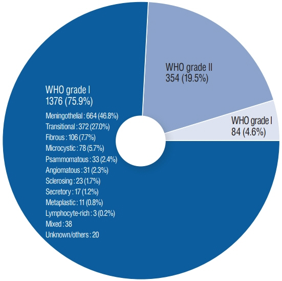
Fig. 1. Distribution of surgically proven meningioma cases from 2004 to 2018 according to WHO grade. There were 1376 (75.9%) WHO grade I benign meningioma cases; 354 (19.5%) and 84 cases (4.6%) were WHO grades II and III, respectively. Among the WHO grade I meningioma, meningothelial subtype was the most common, occurring in 644 of 1376 cases (46.8%). The second most common subtype was transitional meningioma (372 cases, 27.0%), followed by fibrous, microcystic, and psammomatous subtypes. Relatively rare with less than 2% incidence were sclerosing, secretory, metaplastic and lymphocyte-rich meningiomas. Metaplastic meningioma was diagnosed in 11 cases, representing 0.8% of WHO grade I meningioma and 0.6% of all meningiomas during the 15-year study period. The rarest was lymphocyte-rich subtype, with only three cases (0.2%). WHO : World Health Organization.
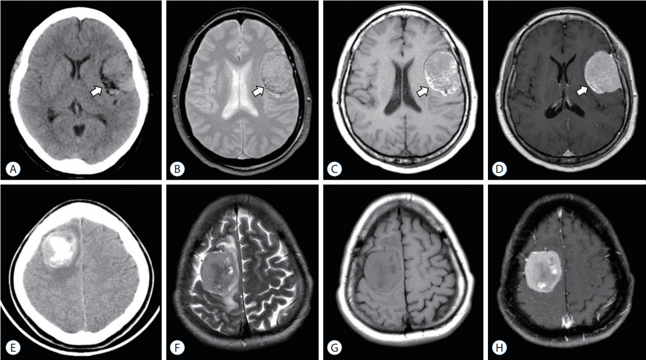
Fig. 2. A-D : Preoperative CT and MR images of lipomatous meningioma (case 1). White arrow suggesting the involvement of fatty components. E-H : Osseous meningioma (case 9). CT : computed tomography, MR : magnetic resonance.

Fig. 3. Gross photo of the surgically resected specimen (formalin-fixed overnight). B : Representative microscopic image shows admixed meningioma tumor cells and fat component (hematoxylin & eosin stain, ×40). C : Membrane immunoreactivity of epithelial membrane antigen immunohistochemical stain observed (×100). D : Ki-67 immunohistochemical stain reveals low proliferative index of tumor cells (Positive in 5%, ×100).
Reference
-
References
1. Barresi V, Caffo M, Ieni A, Alafaci C, Tuccari G. Osteoblastic meningiomas: clinico-pathological and immunohistochemical features of an uncommon variant. J Neurooncol. 105:225–232. 2011.
Article2. Choi YH, Choi CY, Lee CH, Koo HW, Chang SH. Metaplastic meningioma overspreading the cerebral convexity. Brain Tumor Res Treat. 6:97–100. 2018.
Article3. Colnat-Coulbois S, Kremer S, Weinbreck N, Pinelli C, Auque J. Lipomatous meningioma: report of 2 cases and review of the literature. Surg Neurol. 69:398–402. discussion 402. 2008.
Article4. Danisman MC, Kelesoglu KS, Sivri M, Koplay M, Paksoy Y. Microcystic meningioma: difficulties in diagnosis and magnetic resonance imaging findings. Acta Neurol Belg. 117:745–747. 2017.
Article5. Ersoz S, Yilmaz ZS, Eyuboglu I, Yazar U. Xanthomatous meningioma: a case report. Turk Neurosurg. 29:141–144. 2019.
Article6. Gasparinho MG, Ferreira M, Lavrador JP, Livraghi S. Revisiting lipomatous meningioma: a case report and review of an unusual entity. Int J Surg Pathol. 23:399–403. 2015.7. Ikota H, Nakazato Y. A case of metaplastic meningioma with extensive xanthomatous change. Neuropathology. 28:422–426. 2008.
Article8. Indiran V. Intracranial ossified metaplastic meningioma: unusual cause of headache. J Neurosci Rural Pract. 8:653. 2017.
Article9. Ishida M, Fukami T, Nitta N, Iwai M, Yoshida K, Kagotani A, et al. Xanthomatous meningioma: a case report with review of the literature. Int J Clin Exp Pathol. 6:2242–2246. 2013.10. Kang H, Kim JW, Se YB, Dho YS, Choi SH, Park SH. Sclerosing meningioma : radiological and clinical characteristics of 21 cases. J Korean Neurosurg Soc. 59:584–589. 2016.
Article11. Kim NR, Yee GT, Cho HY. Crush cytology of secretory meningioma: a case report. Brain Tumor Res Treat. 3:147–150. 2015.
Article12. Krisht KM, Altay T, Couldwell WT. Myxoid meningioma: a rare metaplastic meningioma variant in a patient presenting with intratumoral hemorrhage. J Neurosurg. 116:861–865. 2012.
Article13. Kunimatsu A, Kunimatsu N, Kamiya K, Katsura M, Mori H, Ohtomo K. Variants of meningiomas: a review of imaging findings and clinical features. Jpn J Radiol. 34:459–469. 2016.
Article14. Mannoji C, Koda M, Murakami M, Kubosawa H, Yamazaki M, Okawa A, et al. Osseous metaplastic meningioma in the thoracic spine mimicking osteosarcoma: a case report. Spine (Phila Pa 1976). 38:E632–E634. 2013.15. Murakami T, Tanishima S, Takeda C, Kato S, Nagashima H. Ossified metaplastic spinal meningioma without psammomatous calcification: a case report. Yonago Acta Med. 62:232–235. 2019.
Article16. Paek SH, Kim SH, Chang KH, Park CK, Kim JE, Kim DG, et al. Microcystic meningiomas: radiological characteristics of 16 cases. Acta Neurochir (Wien). 147:965–972. discussion 972. 2005.17. Salunke P, Aggarwal A, Futane S, Nada R, Gochhait D. Osteoblastic meningioma with turtle shell: different entity from calcified meningioma. Asian J Neurosurg. 11:450. 2016.
Article18. Somerset HL, Kleinschmidt-DeMasters BK, Rubinstein D, Breeze RE. Osteochondroma of the convexity: pathologic-neuroimaging correlates of a lesion that mimics high-grade meningioma. J Neurooncol. 98:421–426. 2010.
Article19. Tang H, Sun H, Chen H, Gong Y, Mao Y, Xie Q, et al. Clinicopathological analysis of metaplastic meningioma: report of 15 cases in Huashan Hospital. Chin J Cancer Res. 25:112–118. 2013.20. Wang DJ, Xie Q, Gong Y, Mao Y, Wang Y, Cheng HX, et al. Histopathological classification and location of consecutively operated meningiomas at a single institution in China from 2001 to 2010. Chin Med J (Engl). 126:488–493. 2013.21. Yüksel MO, Gürbüz MS, Tanriverdi O, Özmen SA. Lipomatous meningioma: a rare subtype of benign metaplastic meningiomas. J Neurosci Rural Pract. 8:140–142. 2017.
Article22. Zouaoui S, Darlix A, Rigau V, Mathieu-Daudé H, Bauchet F, Bessaoud F, et al. Descriptive epidemiology of 13,038 newly diagnosed and histologically confirmed meningiomas in France: 2006-2010. Neurochirurgie. 64:15–21. 2018.
Article
