Comparative observations of lingualized occlusion and monoplane occlusion in the treatment of severe atrophy of edentulous mandible
- Affiliations
-
- 1Department of Prosthodontics, School of Dentistry, Dankook University, Cheonan, Republic of Korea. jseok2@hanmail.net
- KMID: 2422654
- DOI: http://doi.org/10.14368/jdras.2018.34.2.127
Abstract
- The patient who has severely absorbed residual ridges, treatments are challenging to satisfy many factors: support, retention, stability, etc. The neutral zone or monoplane occlusion with non-anatomical tooth would be helpful to get additional retention and stability. The monoplane occlusion has been used long time because it can eliminate horizontal forces and many other advantages. The lingualized occlusion was introduced to improve chewing efficiency and esthetics. But from a stability aspect, it seems controversy between monoplane occlusion and lingualized occlusion. This case report shows the results of the treat two flat residual ridge patients using functional impression; piezography, and made 2 other dentures with monoplane and lingualized occlusion that patient can select denture.
Keyword
Figure
-
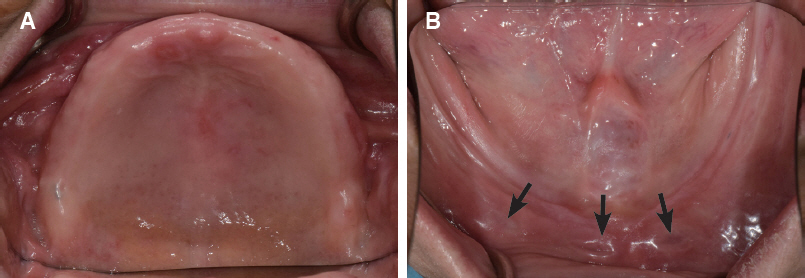
Fig. 1 Initial intra-oral photographs. (A) Maxillary occlusal view, (B) Mandibular occlusal view, severely absorbed ridges observed, arrow: flabby tissue was observed.
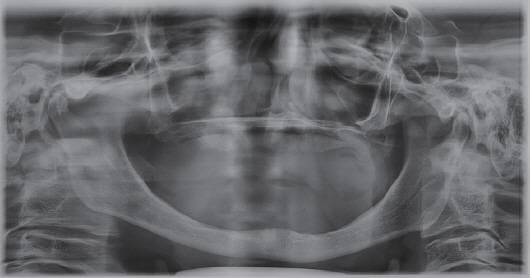
Fig. 2 Panoramic radiograph.
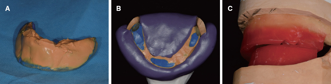
Fig. 3 Taking piezography. (A) After taking piezography by PVS impression material, (B) Putty idex for wax rim, (C) Increased overjet after piezography.
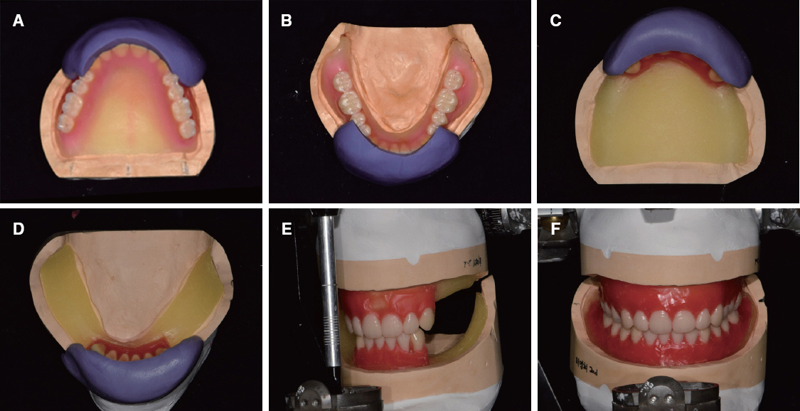
Fig. 4 Denture duplication using putty index. (A) Putty index of upper anterior teeth, (B) Putty index of lower anterior teeth, (C) Arrangement upper anterior teeth by putty index on 2nd denture base, (D) Arrangement lower anterior teeth by putty index on 2nd denture base, (E) After mounting upper and lower cast, (F) Arrangement molar teeth by monoplane occlusion.
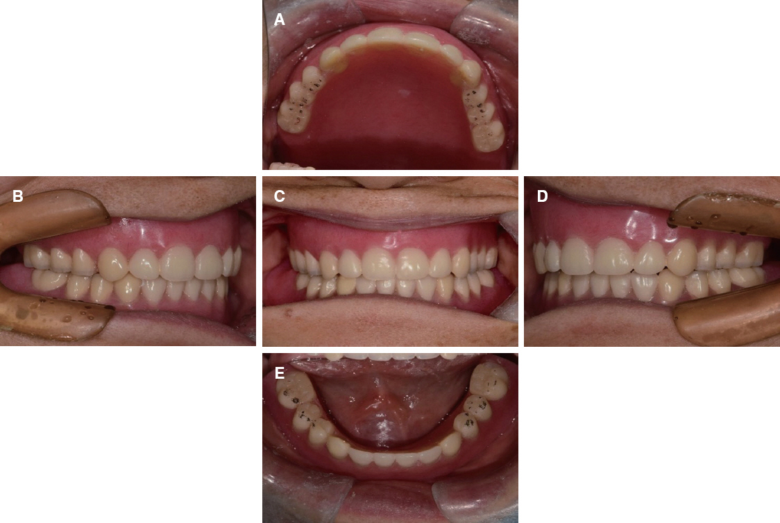
Fig. 5 Monoplane occlusion definitve denture delivery. (A) Maxillary occlusal view, (B) Lateral view (Right), (C) Frontal view, (D) Lateral view (left), (E) Mandibular occlusal view.
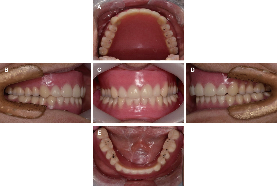
Fig. 6 Lingualized occlusion definitve denture delivery after 3weeks. (A) Maxillary occlusal view, (B) Lateral view (Right), (C) Frontal view, (D) Lateral view (left), (E) Mandibular occlusal view.
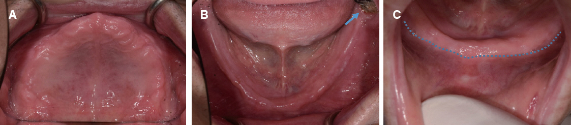
Fig. 7 Initial intraoral photographs. (A) Maxillary occlusal view, (B) Mandibular occlusal view, arrow: Lower left 3rd molar, (C) Lower ridge point on high muscle attachment.
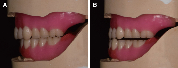
Fig. 8 Arrangement monoplane occlusion with balancing ramps. (A) Lateral view, (B) Tripodism on anterior excursion.
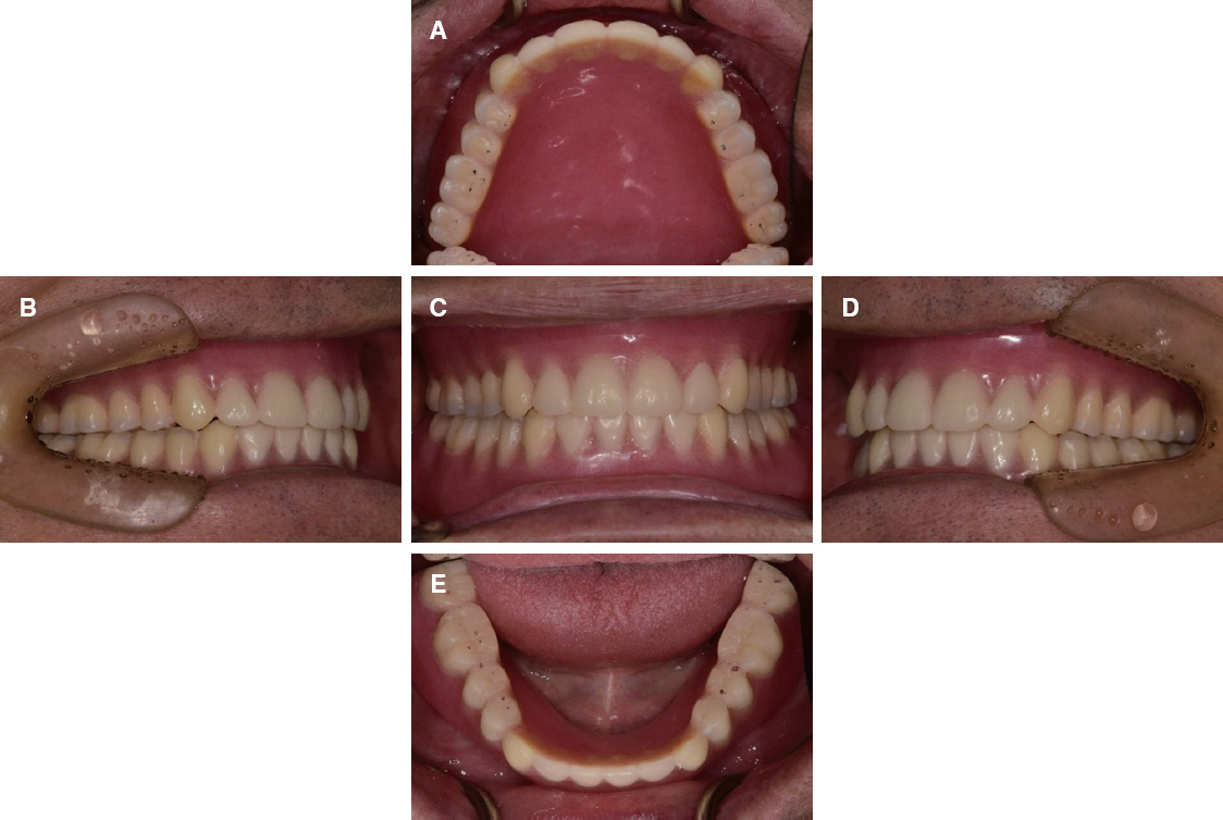
Fig. 9 Lingualized occlusion definitve denture delivery after 3 weeks. (A) Maxillary occlusal view, (B) Lateral view (Right), (C) Frontal view, (D) Lateral view (left), (E) Mandibular occlusal view.

Fig. 10 Monoplane occlusion definitve denture delivery. (A) Maxillary occlusal view, (B) Lateral view (Right), (C) Frontal view, (D) Lateral view (left), (E) Mandibular occlusal view.
Cited by 1 articles
-
The treatment of an edentulous patient with conventional complete denture and CAD/CAM complete denture
Sungyoon Cho, Joonseok Lee
J Korean Acad Prosthodont. 2020;58(1):42-49. doi: 10.4047/jkap.2020.58.1.42.
Reference
-
References
1. Payne SH. A comparative study of posterior occlusion. J Prosthet Dent. 1952; 2:661–6. DOI: 10.1016/S0022-3913(52)80043-5.2. Matsumaru Y. Influence of mandibular residual ridge resorption on objective masticatory measures of lingualized and fully bilateral balanced denture articulation. J Prosthodont Res. 2010; 54:112–8. DOI: 10.1016/j.jpor.2009.11.008. PMID: 20089469.3. Jones PM. The monoplane occlusion for complete dentures. J Am Dent Assoc. 1972; 85:94–100. DOI: 10.14219/jada.archive.1972.0293. PMID: 4503599.4. Phoenix RD, Engelmeier RL. Lingualized occlusion revisited. J Prosthet Dent. 2010; 104:342–6. DOI: 10.1016/S0022-3913(10)60153-9. PMID: 20970541.5. Shetty NS. Comparative observations of the use of cusp and zero-degree posterior teeth. J Prosthet Dent. 1984; 51:459–60. DOI: 10.1016/0022-3913(84)90293-2. PMID: 6374103.6. Berg E. The influence of cusped and cuspless teeth on patient satisfaction with complete dentures. A 2-year follow-up study. J Dent. 1988; 16:269–76. DOI: 10.1016/0300-5712(88)90027-9. PMID: 3065374.7. Nimmo A, Kratochvil FJ. Balancing ramps in nonanatomic complete denture occlusion. J Prosthet Dent. 1985; 53:431–3. DOI: 10.1016/0022-3913(85)90529-3. PMID: 3886890.8. Madalli P, Murali CR, Subhas S, Garg S, Shahi P, Parasher P. Effect of occlusal scheme on the pressure distribution of complete denture supporting tissues:an in vitro study. J Int Oral Health. 2015; 7:68–73. PMID: 26668486. PMCID: PMC4672861.9. Sutton AF, McCord JF. A randomized clinical trial comparing anatomic, lingualized, and zero-degree posterior occlusal forms for complete dentures. J Prosthet Dent. 2007; 97:292–8. DOI: 10.1016/j.prosdent.2007.03.003. PMID: 17547948.10. Ikebe K, Okuno I, Nokubi T. Effect of adding impression material to mandibular denture space in Piezography. J Oral Rehabil. 2006; 33:409–15. DOI: 10.1111/j.1365-2842.2005.01582.x. PMID: 16671986.
- Full Text Links
-
- Actions
-
Cited
- CITED
-
- Close
- Share
-
- Similar articles
-
- Complete denture treatment using lingualized occlusion scheme at the edentulous patient with severely absorbed flat residual ridges: a case report
- Complete denture rehabilitation in the edentulous patient with severe mandibular bone resorption and vascular malformation using closed mouth impression and monoplane occlusion: A case report
- Complete denture rehabilitation of a fully edentulous patient with unilateral facial nerve palsy: A case report
- Full mouth rehabilitation using removable prosthesis of patient with unstable mandible movements: A case report
- A roentgenocephalometric study on the centric occlusion and the rest position of the mandible in the normal occlusion
