Complete denture treatment using lingualized occlusion scheme at the edentulous patient with severely absorbed flat residual ridges: a case report
- Affiliations
-
- 1Department of Prosthodontics, School of Dentistry, Dankook University, Cheonan, Republic of Korea. jseok@hanmail.net
- KMID: 2394644
- DOI: http://doi.org/10.14368/jdras.2017.33.3.207
Abstract
- Many factors should be considered for successful denture treatment at edentulous patients: support, retention, stability, occlusion, esthetics, etc. The patient who has severely absorbed residual ridges, however, treatments are challenging to satisfy those factors. The dentures that use anatomic artificial teeth show good mastication efficiency and esthetics but, can easily lose stability at absorbed ridges. On the contrary, the dentures that use non-anatomic artificial teeth perform better stability but, lower masticatory efficiency and esthetics at absorbed ridges. The lingualized occlusion, using both anatomic and non-anatomic teeth, introduced for compromise those of the pros and cons. At lingualized occlusion, buccal cusps of the teeth do not contact on centric relation. Therefore, direction of the occlusal force towards lingually, then stability of dentures increases. This case report shows the results of the treatment flat residual ridges using complete dentures with ligualized occlusion to increase dentures stability and satisfactory of the patient.
MeSH Terms
Figure
-
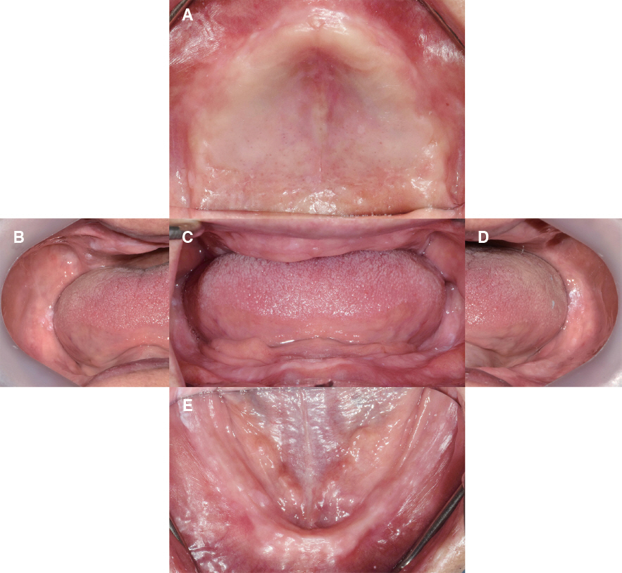
Fig. 1 (A) Upper, (B) right lateral, (C) frontal, (D) left lateral and (E) lower views of intraoral photos at the initial examination. Severely absorbed ridges observed.
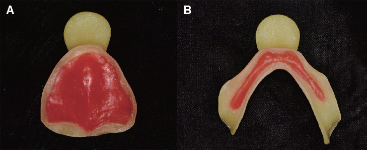
Fig. 2 Individual trays of (A) upper and (B) lower jaws for selective pressure impression technique.
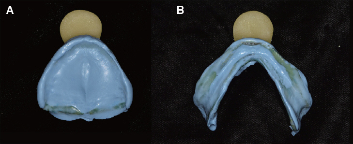
Fig. 3 Impressions of (A) upper and (B) lower jaws that taken by individual trays and addition silicone impression materials.
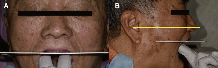
Fig. 4 (A) Frontal and (B) lateral views of occlusal plane setting.
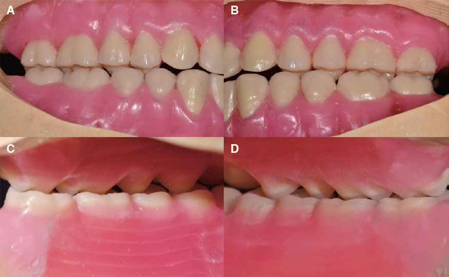
Fig. 5 (A) Right buccal, (B) left buccal, (C) left lingual and (D) right lingual views of wax dentures.
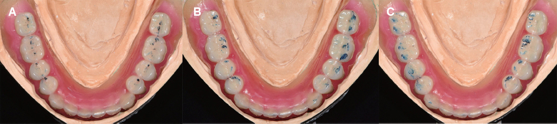
Fig. 6 Occlusal contact point of (A) centric relation, (B) left lateral excursion and (C) right lateral excursion.
Fig. 7 Occlusion registration with alu-wax for clinical remounting.
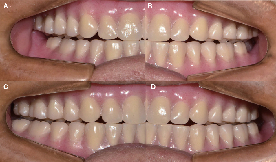
Fig. 8 Confirmation of the lingualized occlusion at intraoral denture delivery phase. The photos of (A) right, (B) left non-working side and (C) right, (D) left working side showed upper palatal cusps evenly contacted to lower molars.
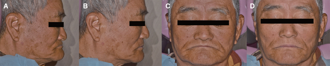
Fig. 9 Lateral and frontal view of extraoral photos of the patient before and after treatment. (B) Lateral and (D) frontal facial profiles were improved esthetically than (A) lateral and (C) frontal facial profiles with old dentures.
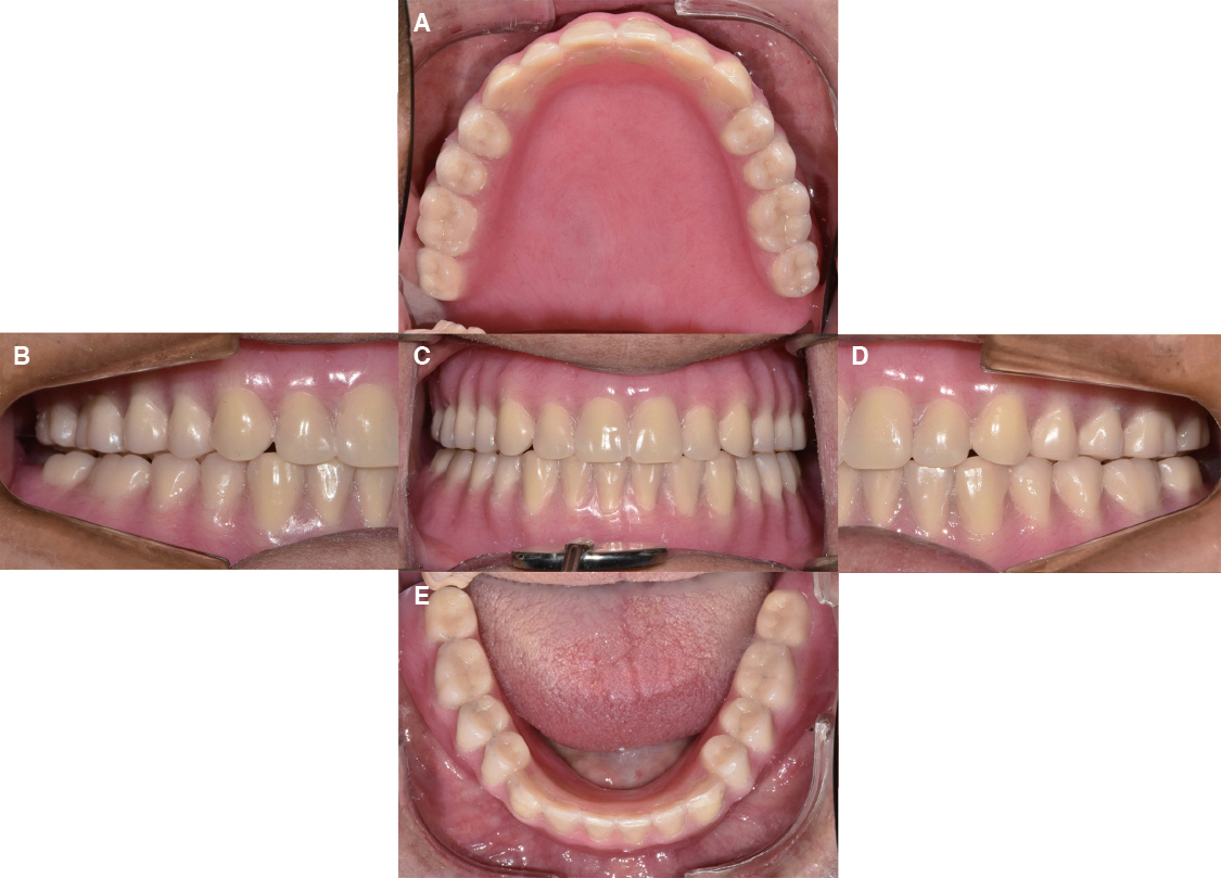
Fig. 10 (A) Upper, (B) right lateral, (C) frontal, (D) left lateral, (E) lower views of the intraoral photos after treatment at centric relation state.
Reference
-
References
1. Kwon WI, Song YG, Lee JS. Complete denture rehabilitation of fully edentulous patient with severe bone resorption and class II jaw relation using piezography. J Korean Acad Prosthodont. 2016; 54:445–50. DOI: 10.4047/jkap.2016.54.4.445.2. Kawai Y, Ikeguchi N, Suzuki A, Kuwashima A, Sakamoto R, Matsumaru Y, Kimoto S, Iijima M, Feine JS. A double blind randomized clinical trial comparing lingualized and fully bilateral balanced posterior occlusion for conventional complete dentures. J Prosthodont Res. 2017; 61:113–22. DOI: 10.1016/j.jpor.2016.07.003. PMID: 27474364.3. Becker CM, Swoope CC, Guckes AD. Lingualized occlusion for removable prosthodontics. J Prosthet Dent. 1977; 38:601–8. DOI: 10.1016/0022-3913(77)90002-6.4. Clough HE, Knodle JM, Leeper SH, Pudwill ML, Taylor DT. A comparison of lingualized occlusion and monoplane occlusion in complete dentures. J Prosthet Dent. 1983; 50:176–9. DOI: 10.1016/0022-3913(83)90007-0.5. Gysi A. Special teeth for cross-bite cases. Dent Digest. 1927; 33:167–71.6. Sutton AF, McCord JF. A randomized clinical trial comparing anatomic, lingualized, and zero-degree posterior occlusal forms for complete dentures. J Prosthet Dent. 2007; 97:292–8. DOI: 10.1016/j.prosdent.2007.03.003. PMID: 17547948.7. Ohnuki M, Nishiyama Y, Hosoi T, Tojo T, Nishimura K. Functional efficacy of full balanced occlusion and lingualized occlusion evaluated from morphological differences in the edentulous alveolar ridge. Prosthodont Res Pract. 2002; 1:31–40. DOI: 10.2186/prp.1.31.8. Matsumaru Y. Influence of mandibular residual ridge resorption on objective masticatory measures of lingualized and fully bilateral balanced denture articulation. J Prosthodont Res. 2010; 54:112–8. DOI: 10.1016/j.jpor.2009.11.008. PMID: 20089469.9. Deniz DA, Kulak Ozkan Y. The influence of occlusion on masticatory performance and satisfaction in complete denture wearers. J Oral Rehabil. 2013; 40:91–8. DOI: 10.1111/joor.12015. PMID: 23189997.10. Heydecke G, Vogeler M, Wolkewitz M, Türp JC, Strub JR. Simplified versus comprehensive fabrication of complete dentures: patient ratings of denture satisfaction from a randomized crossover trial. Quintessence Int. 2008; 39:107–16. PMID: 18560649.11. Zhao K, Mai QQ, Wang XD, Yang W, Zhao L. Occlusal designs on masticatory ability and patient satisfaction with complete denture: a systematic review. J Dent. 2013; 41:1036–42. DOI: 10.1016/j.jdent.2013.07.016. PMID: 23911601.12. Lim SR, Lee JS. Three dimensional deformation of dry-stored complete denture base at room temperature. J Adv Prosthodont. 2016; 8:296–303. DOI: 10.4047/jap.2016.8.4.296. PMID: 27555899. PMCID: PMC4993843.13. Basso MF, Nogueira SS, Arioli-Filho JN. Comparison of the occlusal vertical dimension after processing complete dentures made with lingualized balanced occlusion and conventional balanced occlusion. J Prosthet Dent. 2006; 96:200–4. DOI: 10.1016/j.prosdent.2006.08.005. PMID: 16990071.14. Phoenix RD, Engelmeier RL. Lingualized occlusion revisited. J Prosthet Dent. 2010; 104:342–6. DOI: 10.1016/S0022-3913(10)60153-9.15. Arksornnukit M, Phunthikaphadr T, Takahashi H. Pressure transmission and distribution under denture bases using denture teeth with different materials and cuspal angulations. J Prosthet Dent. 2011; 105:127–36. DOI: 10.1016/S0022-3913(11)60013-9.
- Full Text Links
-
- Actions
-
Cited
- CITED
-
- Close
- Share
-
- Similar articles
-
- Comparative observations of lingualized occlusion and monoplane occlusion in the treatment of severe atrophy of edentulous mandible
- Rehabilitation of a patient with atrophic ridges using gothic arch tracing and nonpressure impression: a case report
- Management of flabby ridges using liquid supported denture: a case report
- Complete denture rehabilitation of fully edentulous patient with severe bone resorption and class II jaw relation using piezography
- A case report on telescopic denture with a small number of residual teeth in mandible
