J Korean Acad Prosthodont.
2017 Oct;55(4):451-457. 10.4047/jkap.2017.55.4.451.
Complete denture rehabilitation of a fully edentulous patient with unilateral facial nerve palsy: A case report
- Affiliations
-
- 1Department of Prosthodontics, Hanil General Hospital, Seoul, Republic of Korea. soonieya@nate.com
- KMID: 2393918
- DOI: http://doi.org/10.4047/jkap.2017.55.4.451
Abstract
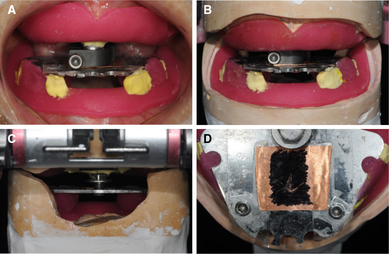
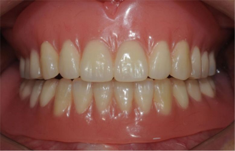
- Bell's palsy is an acute-onset unilateral peripheral facial neuropathy. For patients with sequelae of facial paresis, the successful rehabilitation of fully edentulous arches is challenging. This case report described the treatment procedures and clinical considerations to fabricate complete dentures of a patient who showed unilateral displacement of mandible, unilateral chewing pattern and parafunctional jaw movement due to sequelae of Bell's palsy. Gothic arch tracing was used to record reproducible centric relation and lingualized occlusion was performed to provide freedom to move between centric relation and the patient's habitual functional area in fabricating satisfactory dentures in terms of function and esthetics.
MeSH Terms
Figure
Reference
-
1.Eviston TJ., Croxson GR., Kennedy PG., Hadlock T., Krishnan AV. Bell's palsy: aetiology, clinical features and multidisciplinary care. J Neurol Neurosurg Psychiatry. 2015. 86:1356–61.
Article2.Dawson PE. Centric relation. Its effect on occluso-muscle harmony. Dent Clin North Am. 1979. 23:169–80.3.Payne SH. A posterior set-up to meet individual requirements. Dent Digest. 1941. 47:20–2.4.Lang BR. Complete denture occlusion. Dent Clin North Am. 1996. 40:85–101.
Article5.House JW., Brackmann DE. Facial nerve grading system. Otolaryngol Head Neck Surg. 1985. 93:146–7.
Article6.Carlsson GE. Symptoms of mandibular dysfunction in complete denture wearers. J Dent. 1976. 4:265–70.
Article7.Mercado MD., Faulkner KD. The prevalence of craniomandibular disorders in completely edentulous denture-wearing subjects. J Oral Rehabil. 1991. 18:231–42.
Article8.Chen CK., Tang YB. Myectomy and botulinum toxin for paralysis of the marginal mandibular branch of the facial nerve: a series of 76 cases. Plast Reconstr Surg. 2007. 120:1859–64.
Article9.Yurkstas AA., Kapur KK. Factors influencing centric relation records in edentulous mouths. 1964. J Prosthet Dent. 2005. 93:305–10.10.Smith HF Jr. A comparison of empirical centric relation records with location of terminal hinge axis and apex of the gothic arch tracing. J Prosthet Dent. 1975. 33:511–20.
Article11.Myers M., Dziejma R., Goldberg J., Ross R., Sharry J. Relation of Gothic arch apex to dentist-assisted centric relation. J Prosthet Dent. 1980. 44:78–81.
Article12.Utz KH., Müller F., Bernard N., Hültenschmidt R., Kurbel R. Comparative studies on check-bite and central-bearing-point method for the remounting of complete dentures. J Oral Rehabil. 1995. 22:717–26.
Article13.Linsen SS., Stark H., Samai A. The influence of different registration techniques on condyle displacement and electromyographic activity in stomatognathically healthy subjects: a prospective study. J Prosthet Dent. 2012. 107:47–54.
Article14.Becker CM., Swoope CC., Guckes AD. Lingualized occlusion for removable prosthodontics. J Prosthet Dent. 1977. 38:601–8.
Article15.Phoenix RD., Engelmeier RL. Lingualized occlusion revisited. J Prosthet Dent. 2010. 104:342–6.
Article16.Inoue S., Kawano F., Nagao K., Matsumoto N. An in vitro study of the influence of occlusal scheme on the pressure distribution of complete denture supporting tissues. Int J Prosthodont. 1996. 9:179–87.
- Full Text Links
- Actions
-
Cited
- CITED
-
- Close
- Share
- Similar articles
-
- Comparative analysis of case series for the prosthetic rehabilitation of edentulous patients using suction denture
- Comparison of conventional and suction-effective complete denture in a fully edentulous patient: a case report
- Approach to complicated fully edentulous case: from the diagnosis to the definitive denture
- Suction-effective complete denture fabrication of edentulous patient using BPS: a case report
- The Implant Retained Overdenture by Locator Attachments on the Edentulous Mandible: A Case Report


