Postoperative Chylothorax: the Use of Dynamic Magnetic Resonance Lymphangiography and Thoracic Duct Embolization
- Affiliations
-
- 1Department of Radiology and Research Institute of Radiology, Asan Medical Center, Seoul, Korea. radkoo@amc.seoul.kr
- KMID: 2421551
- DOI: http://doi.org/10.13104/imri.2018.22.3.182
Abstract
- Dynamic enhanced magnetic resonance lymphangiography can be used to provide anatomic and dynamic information for various lymphatic diseases, including thoracic duct injury, and can also help to guide the thoracic duct embolization procedure. We present a case of postoperative chylothorax demonstrated by dynamic enhanced MR lymphangiography. In this case, the chyle leakage site and location of cisterna chyli were clearly visualized by dynamic enhanced MR lymphangiography, thus allowing for management with thoracic duct embolization.
Keyword
Figure
-
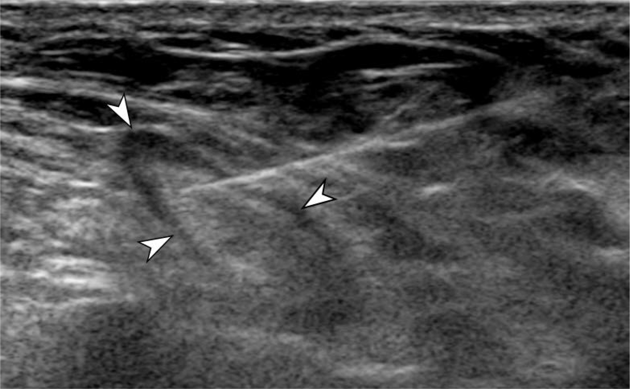
Fig. 1. Ultrasound-guided inguinal lymph node (arrowheads) selection.
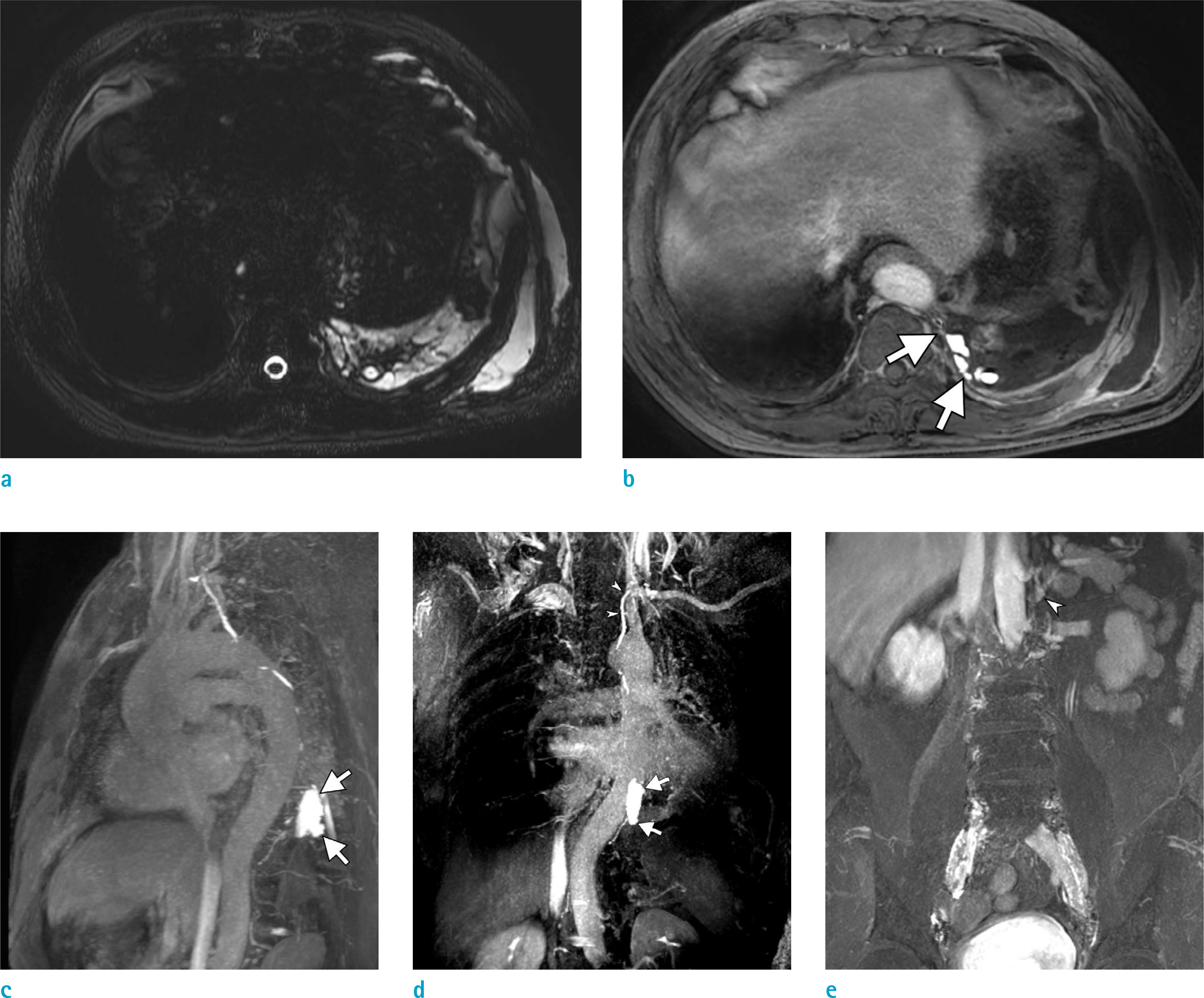
Fig. 2. MR lymphangiography for visualizing the cisterna chyli and leakage site. (a) A T2W image showing postoperative multiloculated fluid collection in the left pleural space and chest wall. (b) An enhanced 3D THRIVE image and (c, d) volume-rendered image with the enhanced 3D THRIVE sequence (20 mm thickness) showing the leakage site (arrows) and that the thoracic duct drains into left subclavian vein (arrowheads). (e) Cisterna chyli (arrowhead) was noted on the left side of the T12 level.
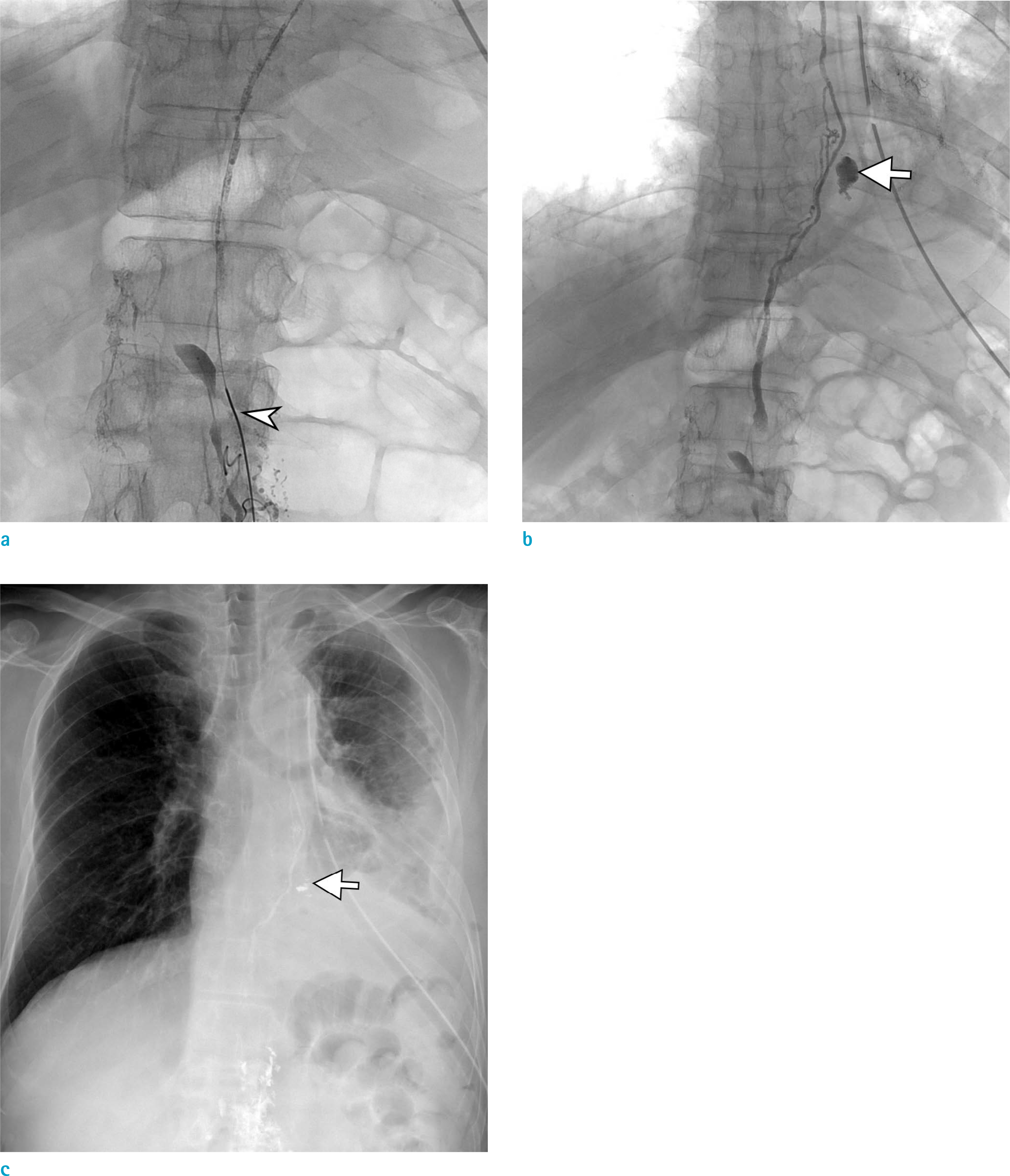
Fig. 3. Conventional lymphangiography with thoracic duct embolization. (a) The cisterna chyli (arrowhead) was selected using a 2.0 Fr Progreat catheter, and Lipiodol uptake was visualized along the thoracic duct course. (b) Thoracic duct embolization was performed at the leakage site (arrow) using histoacryl glue. (c) Immediate chest radiography revealed the embolized leakage site (arrow) and thoracic duct.
Reference
-
References
1. Erden A, Fitoz S, Yagmurlu B, Erden I. Abdominal confluence of lymph trunks: detectability and morphology on heavily T2-weighted images. AJR Am J Roentgenol. 2005; 184:35–40.
Article2. Deso S, Ludwig B, Kabutey NK, Kim D, Guermazi A. Lymphangiography in the diagnosis and localization of various chyle leaks. Cardiovasc Intervent Radiol. 2012; 35:117–126.
Article3. Krishnamurthy R, Hernandez A, Kavuk S, Annam A, Pimpalwar S. Imaging the central conducting lymphatics: initial experience with dynamic MR lymphangiography. Radiology. 2015; 274:871–878.
Article4. Lohrmann C, Foeldi E, Speck O, Langer M. High-resolution MR lymphangiography in patients with primary and secondary lymphedema. AJR Am J Roentgenol. 2006; 187:556–561.
Article5. Pillay TG, Singh B. A review of traumatic chylothorax. Injury. 2016; 47:545–550.
Article
- Full Text Links
-
- Actions
-
Cited
- CITED
-
- Close
- Share
-
- Similar articles
-
- Lymphangiography to Treat Postoperative Lymphatic Leakage: A Technical Review
- Thoracic Duct Embolization with Lipiodol for Chylothorax due to Thoracic Endovascular Aortic Repair with Debranching Procedure
- Interventional Radiology Treatment for Postoperative Chylothorax
- Single-Center Experience With Dynamic Contrast-Enhanced Magnetic Resonance Lymphangiography for Diagnosing Lymphatic Disorders and Guiding Percutaneous Embolization
- Novel Interventional Radiology for the Treatment of Various Lymphatic Leakages: Lymphatic Intervention and Embolization
