Histomorphometric demonstration of the effect of chronic use of nonsteroidal anti-inflammatory drugs–ibuprofen on mucosa of small intestine
- Affiliations
-
- 1Department of Anatomy, People's College of Medical Sciences, Bhopal, India. dendoctor5@gmail.com
- 2Department of Anatomy, MGIMS, Sewagram, India.
- KMID: 2421175
- DOI: http://doi.org/10.5115/acb.2018.51.2.113
Abstract
- The purpose of this study was to ascertain change in structure of mucosa of small intestine, if any, in small intestine of Swiss albino mice as an effect of chronic use of nonsteroidal anti-inflammatory drugs-Ibuprofen. Longitudinal study conducted on 46 adult Swiss albino mice, 23 as experimental and 23 as control. Ibuprofen was given at a dose of 40 µg/g body weight per day for 6 weeks by intragastric route in experimental group of mice while control group of mice received same volume of distilled water. Mice of both the groups were sacrificed and desired segments of small intestines were dissected out and tissues were subjected to histological processing. Histomorphometry was performed and relevant photomicrographs were obtained. Student's unpaired t test by GraphPad Prism 6 software. Height of villi was not significantly altered but there was significant reduction of the number of goblet and non-goblet cells (enterocytes and other columnar cells) in mucosal lining of the small intestine of experimental group of mice. Percent distribution of the goblet and non-goblet cells was not altered in villi of two groups. Chronic exposure of Ibuprofen in therapeutic dosage caused reduction of the functional cell mass in lining epithelium of villi of middle segment of small intestine. However, there was no evidence of ulcerative or hemorrhagic lesion.
Keyword
MeSH Terms
Figure
-
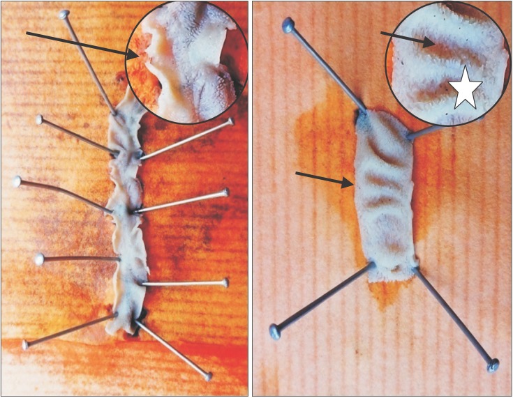
Fig. 1 Gross examination of the interior of intestinal segment: note normal appearance of plicae circulares (*) and ‘villi’ as furry coat (arrows).

Fig. 2 Distribution of goblet and non-goblet cells per villus (%) in control group shown by pie diagram.
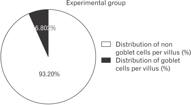
Fig. 3 Distribution of goblet and non-goblet cells per villus (%) in experimental group shown by pie diagram.

Fig. 4 Section of small intestine showing distribution (arrows) of goblet cells (control group; Alcian blue, ×100).

Fig. 5 Section of small intestine showing distribution (arrows) of goblet cells (experimental group; Alcian blue, ×100).

Fig. 6 Section of small intestine showing goblet cell (G), enterocytes (E), and villus core (*) (control; H&E, 1,000).
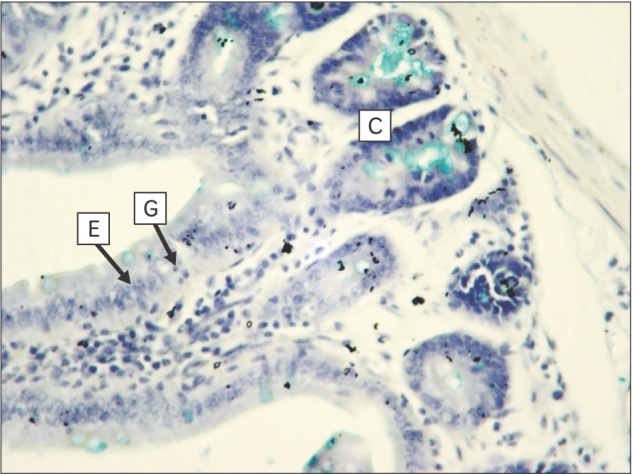
Fig. 7 Section of small intestine showing goblet cells (G), enterocytes (E) on villus with villus core (*) and crypt (C) (experimental; Alcian blue, ×400).
Reference
-
1. Tacheci I, Kopacova M, Rejchrt S, Bures J. Non-steroidal anti-inflammatory drug induced injury to the small intestine. Acta Medica (Hradec Kralove). 2010; 53:3–11. PMID: 20608226.2. Smale S, Tibble J, Sigthorsson G, Bjarnason I. Epidemiology and differential diagnosis of NSAID-induced injury to the mucosa of the small intestine. Best Pract Res Clin Gastroenterol. 2001; 15:723–738. PMID: 11566037.3. Chatterjee TK. Handbook of laboratory mice and rats. Calcutta: K.K. Chatterjee Publication;1993. p. 3–12.4. Drury RA, Wallington EA. Carleton's histological technique. 5th ed. New York: Oxford University Press;1980. p. 57–75.5. Ettarh RR, Carr KE. Morphometric analysis of the small intestinal epithelium in the indomethacin-treated mouse. J Anat. 1996; 189(Pt 1):51–56. PMID: 8771395.6. Anthony A, Dhillon AP, Nygard G, Hudson M, Piasecki C, Strong P, Trevethick MA, Clayton NM, Jordan CC, Pounder RE, Wakefield AJ. Early histological features of small intestinal injury induced by indomethacin. Aliment Pharmacol Ther. 1993; 7:29–39. PMID: 8439635.7. Kuroda M, Yoshida N, Ichikawa H, Takagi T, Okuda T, Naito Y, Okanoue T, Yoshikawa T. Lansoprazole, a proton pump inhibitor, reduces the severity of indomethacin-induced rat enteritis. Int J Mol Med. 2006; 17:89–93. PMID: 16328016.8. Menozzi A, Pozzoli C, Giovannini E, Solenghi E, Grandi D, Bonardi S, Bertini S, Vasina V, Coruzzi G. Intestinal effects of non-selective and selective cyclooxygenase inhibitors in the rat. Eur J Pharmacol. 2006; 552:143–150. PMID: 17069793.9. Dixon JM, Paulley JW. Bacteriological and histological studies of the small intestine of rats treated with mecamylamine. Gut. 1963; 4:169–173. PMID: 14028129.10. Fortun PJ, Hawkey CJ. Nonsteroidal antiinflammatory drugs and the small intestine. Curr Opin Gastroenterol. 2007; 23:134–141. PMID: 17268241.
- Full Text Links
-
- Actions
-
Cited
- CITED
-
- Close
- Share
-
- Similar articles
-
- A Case of Eosinophilic Pneumonia with Ibuprofen as the Suspected Etiology
- Endoscopic Hemostasis for Bleeding Gastric Ulcer Caused by Ibuprofen in a 16-month-old Infant
- A Case of Acute Generalized Exanthematous Pustulosis
- A Study on the Effect of Topical Nonsteroidal Anti - inflammatory Drugs And Cortisosteroids on Ultraviolet Light - Induced Erythema
- A Case of Ischemic Colitis Related with Usual Dosage of Ibuprofen in a Young Man
