Korean J Gastroenterol.
2018 Sep;72(3):150-154. 10.4166/kjg.2018.72.3.150.
Primary Hepatic Schwannoma
- Affiliations
-
- 1Department of Internal Medicine, Gachon University Gil Medical Center, Incheon, Korea. jhkim@gilhospital.com
- 2Department of Radiology, Gachon University Gil Medical Center, Incheon, Korea.
- 3Department of Pathology, Gachon University Gil Medical Center, Incheon, Korea.
- KMID: 2420754
- DOI: http://doi.org/10.4166/kjg.2018.72.3.150
Abstract
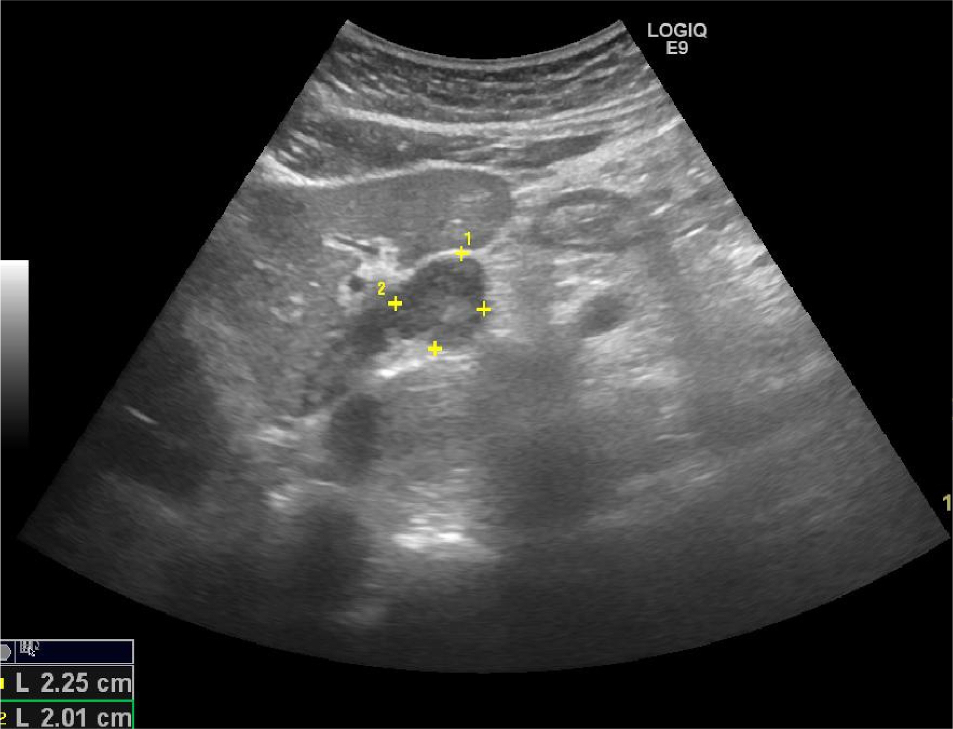
- A primary benign schwannoma of the liver is extremely rare. Only 30 cases have been reported in the medical literature worldwide, and only one case has been reported in Korea previously. A 56-year-old man was admitted to Gil Medical Center with incidental findings of a hepatic mass by abdominal computed tomography. The computed tomography and magnetic resonance image revealed a 3×2 cm-sized solid mass in the left lobe of the liver. Histological examination confirmed the diagnosis of a benign schwannoma, proven by positive immunoreaction with the neurogenic marker S-100 protein and a negative response to CD34, CD117, and smooth muscle actin. We report a primary benign schwannoma of the liver and review the literature.
MeSH Terms
Figure
Reference
-
References
1. Albert P, Patel J, Badawy K, et al. Peripheral nerve schwannoma: a review of varying clinical presentations and imaging findings. J Foot Ankle Surg. 2017; 56:632–637.
Article2. Yamamoto M, Hasegawa K, Arita J, et al. Primary hepatic schwannoma: a case report. Int J Surg Case Rep. 2016; 29:146–150.
Article3. Jung HI, Lee HU, Ahn TS, et al. Primary hepatic malignant peripheral nerve sheath tumor successfully treated with combination therapy: a case report and literature review. Ann Surg Treat Res. 2016; 91:327–331.
Article4. Yin SY, Zhai ZL, Ren KW, et al. Porta hepatic schwannoma: case report and a 30-year review of the literature yielding 15 cases. World J Surg Oncol. 2016; 14:103.
Article5. Momtahen AJ, Akduman EI, Balci NC, Fattahi R, Havlioglu N. Liver schwannoma: findings on MRI. Magn Reson Imaging. 2008; 26:1442–1445.
Article6. Young SJ. Primary malignant neurilemmoma (schwannoma) of the liver in a case of neurofibromatosis. J Pathol. 1975; 117:151–153.
Article7. Lee WH, Kim TH, You SS, et al. Benign schwannoma of the liver: a case report. J Korean Med Sci. 2008; 23:727–730.
Article8. Shih YC, Chen YL, Fang HY, Wu CY, Lin YC, Lin YM. Schwannoma mimicking liver tumor. Thorac Cardiovasc Surg. 2009; 57:436–439.
Article9. Ozkan EE, Guldur ME, Uzunkoy A. A case report of benign schwannoma of the liver. Intern Med. 2010; 49:1533–1536.
Article10. Madhusudhan KS, Srivastava DN, Dash NR, Gupta C, Gupta SD. Case report. Schwannoma of both intrahepatic and extrahepatic bile ducts: a rare case. Br J Radiol. 2009; 82:e212–e215.11. Kulkarni N, Andrews SJ, Rao V, Rajagopal KV. Case report: benign porta hepatic schwannoma. Indian J Radiol Imaging. 2009; 19:213–215.
Article12. Yoshida M, Nakashima Y, Tanaka A, Mori K, Yamaoka Y. Benign schwannoma of the liver: a case report. Nihon Geka Hokan. 1994; 63:208–214.13. Kim YC, Park MS. Primary hepatic schwannoma mimicking malignancy on fluorine-18 2-fluoro-2-deoxy-D-glucose positron emission tomography-computed tomography. Hepatology. 2010; 51:1080–1081.
Article14. Hayashi M, Takeshita A, Yamamoto K, Tanigawa N. Primary hepatic benign schwannoma. World J Gastrointest Surg. 2012; 4:73–78.
Article15. Ota Y, Aso K, Watanabe K, et al. Hepatic schwannoma: imaging findings on CT, MRI and contrastenhanced ultrasonography. World J Gastroenterol. 2012; 18:4967–4972.
Article16. Lederman SM, Martin EC, Laffey KT, Lefkowitch JH. Hepatic neurofibromatosis, malignant schwannoma, and angiosarcoma in von Recklinghausen's disease. Gastroenterology. 1987; 92:234–239.
Article17. Heffron TG, Coventry S, Bedendo F, Baker A. Resection of primary schwannoma of the liver not associated with neurofibromatosis. Arch Surg. 1993; 128:1396–1398.
Article18. Godlewski G, Sawan S, Targhetta P, Pignodel C, Marty-Double C, Gaujoux AF. A malignant schwannoma of the jejunum associated with multiple neurofibromas and a primary adenoma of the parathyroid. Ann Gastroenterol Hepatol (Paris). 1989; 25:13–17.19. Ikehara H, Li Z, Watari J, et al. Histological diagnosis of gastric submucosal tumors: a pilot study of endoscopic ultrasonography-guided fine-needle aspiration biopsy vs mucosal cutting biopsy. World J Gastrointest Endosc. 2015; 7:1142–1149.
Article20. Harwalkar JA, Lee JH, Hughes G, Kinney SE, Golubić M. Immunoblotting analysis of schwannomin/merlin in human schwannomas. Am J Otol. 1998; 19:654–659.


