J Korean Ophthalmol Soc.
2018 Sep;59(9):861-866. 10.3341/jkos.2018.59.9.861.
Poorly Differentiated Neuroendocrine Carcinoma of the Eyelid
- Affiliations
-
- 1Department of Ophthalmology, Samsung Medical Center, Sungkyunkwan University School of Medicine, Seoul, Korea. ydkimoph@skku.edu
- 2Department of Pathology, Samsung Medical Center, Sungkyunkwan University School of Medicine, Seoul, Korea.
- KMID: 2420335
- DOI: http://doi.org/10.3341/jkos.2018.59.9.861
Abstract
- PURPOSE
To report a case of poorly differentiated neuroendocrine carcinoma of the eyelid.
CASE SUMMARY
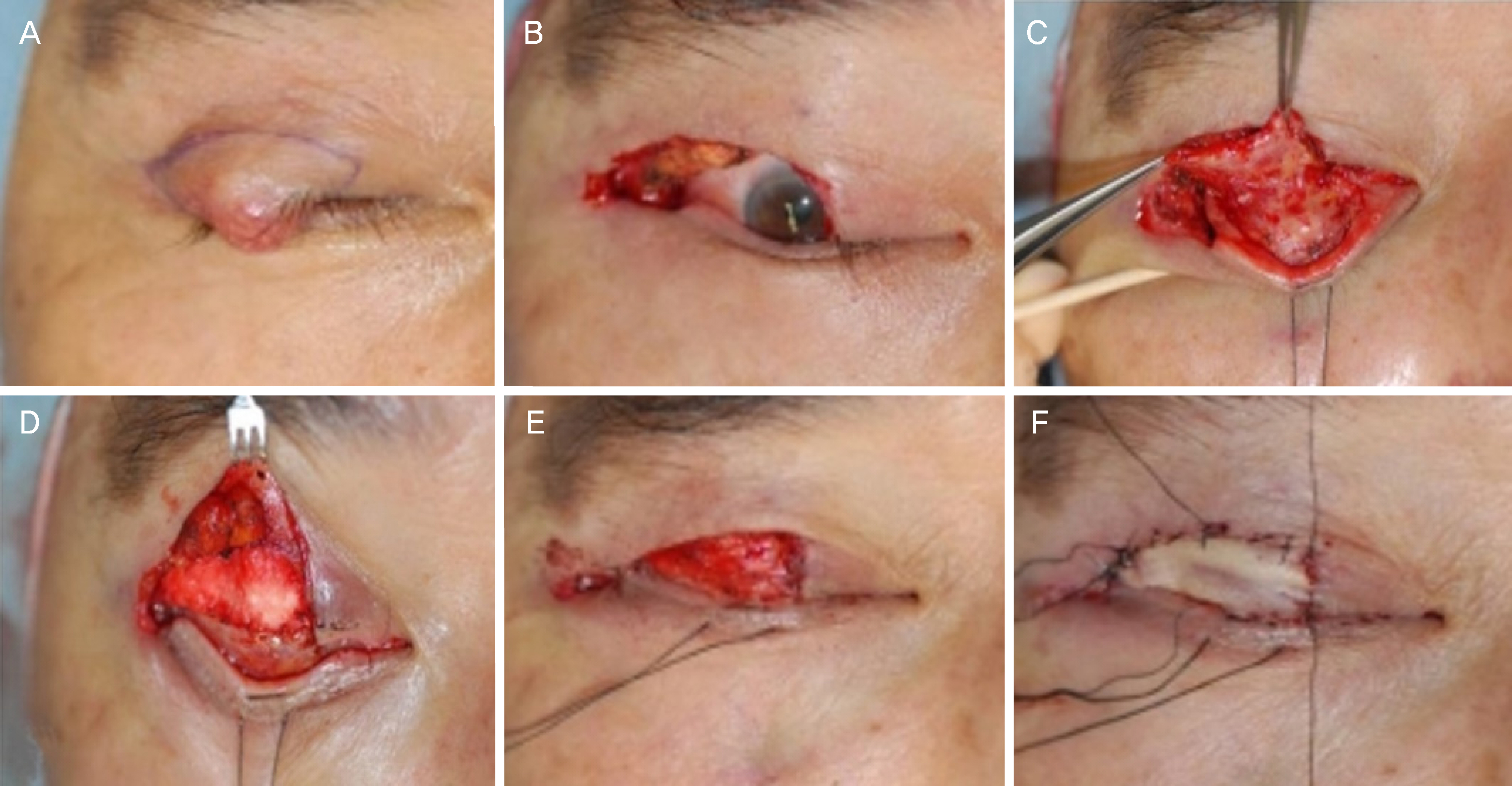
A 70-year-old male presented with a 5-month history of a right upper eyelid mass. The mass appeared as 1.2 × 1.2 cm on the right upper eyelid. A mass excision was performed under frozen section control. The tumor was completely excised with a safety margin clearance and an upper eyelid reconstruction was performed. Histopathological examination revealed a tumor composed of small atypical cells which showed a high nuclear/cytoplasm ratio, nuclear molding, and increased mitotic activity. Immunohistochemical examination revealed positive reactivity for Ki-67, synaptophysin, CD56, and negative reactivity for chromogranin, cytokeratin 20, and thyroid transcription factor-1.
CONCLUSIONS
Primary neuroendocrine carcinoma of the eyelid is extremely rare, but the tumor has high malignancy and readily metastasizes. Poorly differentiated neuroendocrine carcinoma should be considered in the differential diagnosis of a rapidly growing eyelid mass.
Keyword
MeSH Terms
Figure
Reference
-
References
1. Remick SC, Hafez GR, Carbone PP. Extrapulmonary small-cell carcinoma. A review of the literature with emphasis on therapy and outcome. Medicine (Baltimore). 1987; 66:457–71.2. Yao JC, Hassan M, Phan A, et al. One hundred years after “carcinoid”: epidemiology of and prognostic factors for abdominal tumors in 35,825 cases in the United States. J Clin Oncol. 2008; 26:3063–72.3. Bajetta E, Catena L, Procopio G, et al. Is the new WHO classi fi abdominal of neuroendocrine tumours useful for selecting an appropriate treatment? Ann Oncol. 2005; 16:1374–80.4. Solcia EKG, Sobin LH. WHO: Histological Typing of Endocrine Tumors. Berlin/New York: Springer;2000. p. 7–13.5. Faggiano A, Mansueto G, Ferolla P, et al. Diagnostic and abdominal implications of the World Health Organization classi fi cation of neuroendocrine tumors. J Endocrinol Invest. 2008; 31:216–23.6. Remick SC, Ruckdeschel JC. Extrapulmonary and pulmonary small-cell carcinoma: tumor biology, therapy, and outcome. Med Pediatr Oncol. 1992; 20:89–99.
Article7. Galanis E, Frytak S, Lloyd RV. Extrapulmonary small cell carcinoma. Cancer. 1997; 79:1729–36.
Article8. Silkiss RZ, Green JE, Shetlar DJ. Small cell neuroendocrine abdominal of the eyelid. Ophthal Plast Reconstr Surg. 2008; 24:319–21. discussion 321–2.9. Yamanouchi D, Oshitari T, Nakamura Y, et al. Primary abdominal carcinoma of ocular adnexa. Case Rep Ophthalmol Med. 2013; 2013:281351.10. Metz KA, Jacob M, Schmidt U, et al. Merkel cell carcinoma of the eyelid: histological and immunohistochemical features with abdominal respect to differential diagnosis. Graefes Arch Clin Exp Ophthalmol. 1998; 236:561–6.11. Llombart B, Monteagudo C, López-Guerrero JA, et al. Clinicopathological and immunohistochemical analysis of 20 cases of Merkel cell carcinoma in search of prognostic markers. Histopathology. 2005; 46:622–34.
Article12. Hanly AJ, Elgart GW, Jorda M, et al. Analysis of thyroid transcription factor-1 and cytokeratin 20 separates merkel cell carcinoma from small cell carcinoma of lung. J Cutan Pathol. 2000; 27:118–20.
Article13. Swann MH, Yoon J. Merkel cell carcinoma. Semin Oncol. 2007; 34:51–6.
Article14. Dancey AL, Rayatt SS, Soon C, et al. Merkel cell carcinoma: a abdominal of 34 cases and literature review. J Plast Reconstr Aesthet Surg. 2006; 59:1294–9.15. Muqit MM, Roberts F, Lee WR, Kemp E. Improved survival rates in sebaceous carcinoma of the eyelid. Eye (Lond). 2004; 18:49–53.
Article16. Jabbour J, Cumming R, Scolyer RA, et al. Merkel cell carcinoma: assessing the effect of wide local excision, lymph node dissection, and radiotherapy on recurrence and survival in early-stage abdominal–results from a review of 82 consecutive cases diagnosed abdominal 1992 and 2004. Ann Surg Oncol. 2007; 14:1943–52.17. Ho VH, Ross MI, Prieto VG, et al. Sentinel lymph node biopsy for sebaceous cell carcinoma and melanoma of the ocular adnexa. Arch Otolaryngol Head Neck Surg. 2007; 133:820–6.
Article
- Full Text Links
- Actions
-
Cited
- CITED
-
- Close
- Share
- Similar articles
-
- Moderately Differentiated Neuroendocrine Carcinoma of the Larynx
- Two Cases of Merkel Cell Carcinoma of Eyelid and Neck
- Skin Metastasis of Neuroendocrine Carcinoma Arising in the Rectum
- Primary Small Cell Neuroendocrine Carcinoma of the Breast: A Case Report With Literature Review
- Combined Hepatocellular Carcinoma and Neuroendocrine Carcinoma with Ectopic Secretion of Parathyroid Hormone: A Case Report and Review of the Literature


