Ann Rehabil Med.
2018 Aug;42(4):617-620. 10.5535/arm.2018.42.4.617.
Which Neural Tract Plays a Major Role in Memory Impairment After Multiple Cerebral Infarcts? A Case Report
- Affiliations
-
- 1Department of Physical Medicine and Rehabilitation, Ulsan University Hospital, University of Ulsan College of Medicine, Ulsan, Korea. fnew1@hanmail.net
- KMID: 2420055
- DOI: http://doi.org/10.5535/arm.2018.42.4.617
Abstract
- Injury to the thalamocortical tract (one in the Papez circuit) that leads to memory impairment following brain injury is very rare. In this study, we present a case of partial injury to the thalamocortical tract that causes memory impairment after concurrent thalamic and hippocampal infarct. A 20-year-old male complained of memory impairment 1 month after partial injury to the thalamocortical tract. Using a probabilistic diffusing tensor tractography, it was found that the right thalamocortical tract was thinner than the left thalamocortical tract. However, all other neural tracts including the fornix, cingulum, and mammillothalamic tract were intact on both hemispheres. Therefore, the memory impairment in this patient was considered as being due to thalamic infarct based on the observation that the fornix from hippocampal infarct was intact. This case suggests that the assessment of lesions in the neural tracts of the Papez circuit might be useful for understanding the mechanism of memory impairment following cerebral infarction.
Keyword
MeSH Terms
Figure
-
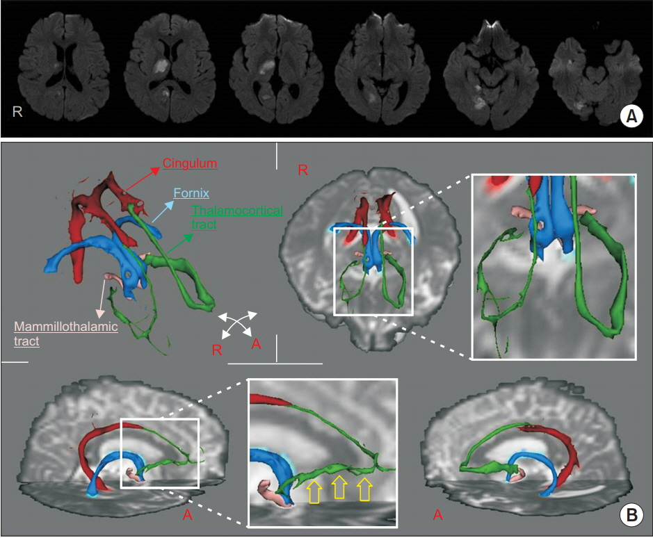
Fig. 1. (A) Diffusion brain magnetic resonance imaging showed cerebral infarction in the anterior thalamus, hippocampus, and the partially fusiform area on the right hemisphere. (B) The right thalamocortical tract (green color) between the anterior thalamic nuclei and the cingulate gyrus is thinner compared to the left thalamocortical tract (yellow arrow). Other tracts such as the mammillothalamic tract (pink), fornix (blue), and cingulum (red) are well constructed without abnormality. R, right; A, anterior.
Reference
-
1. Snaphaan L, de Leeuw FE. Poststroke memory function in nondemented patients: a systematic review on frequency and neuroimaging correlates. Stroke. 2007; 38:198–203.2. Kumral E, Deveci EE, Erdogan C, Enustun C. Isolated hippocampal infarcts: vascular and neuropsychological findings. J Neurol Sci. 2015; 356:83–9.
Article3. Carlesimo GA, Lombardi MG, Caltagirone C. Vascular thalamic amnesia: a reappraisal. Neuropsychologia. 2011; 49:777–89.
Article4. Mori S, Aggarwal M. In vivo magnetic resonance imaging of the human limbic white matter. Front Aging Neurosci. 2014; 6:321.
Article5. Jang SH, Kwon HG. Diffuse injury of the Papez circuit by focal head trauma: a diffusion tensor tractography study. Acta Neurol Belg. 2017; 117:389–91.
Article6. Chang MC, Yeo SS, Do Lee H, Jang SH. Diffusion tensor tractography in a patient with memory impairment following encephalitis. Acta Neurol Belg. 2016; 116:629–31.
Article7. Jang SH, Yeo SS. Injury of the Papez circuit in a patient with provoked confabulation following subarachnoid hemorrhage: a diffusion tensor tractography study. Acta Neurol Belg. 2016; 116:655–8.
Article8. Yang DS, Kwon HG, Jang SH. Injury of the thalamocingulate tract in the papez circuit in patients with mild traumatic brain injury. Am J Phys Med Rehabil. 2016; 95:e34. –8.
Article9. Kwon HG, Chang CH, Jang SH. Is thalamocortical tract injury responsible for memory impairment in a patient with putaminal hemorrhage? Neural Regen Res. 2015; 10:321–2.
Article10. Kwon HG, Lee HD, Jang SH. Injury of the mammillothalamic tract in patients with thalamic hemorrhage. Front Hum Neurosci. 2014; 8:259.
Article
- Full Text Links
-
- Actions
-
Cited
- CITED
-
- Close
- Share
-
- Similar articles
-
- Possible Role of a Missense Mutation of p.P167S on NOTCH3 Gene Associated with Cerebral Autosomal Dominant Arteriopathy with Subcortical Infarcts and Leukoencephalopathy
- Hypereosinophilia with Multiple Thromboembolic Cerebral Infarcts and Focal Intracerebral Hemorrhage
- Importance of Symptomatic Cerebral Infarcts on Cognitive Performance in Patients with Alzheimer's Disease
- Memory Impairment in Dementing Patients
- Effect of Synaptic Loss on Memory in a Neural Network Model
