Clin Endosc.
2018 Jul;51(4):329-333. 10.5946/ce.2018.095.
Current Status of Interpretation of Small Bowel Capsule Endoscopy
- Affiliations
-
- 1Department of Internal Medicine, Seoul Metropolitan Government Seoul National University Boramae Medical Center, Seoul National University College of Medicine, Seoul, Korea.
- 2Department of Gastroenterology, Asan Medical Center, University of Ulsan College of Medicine, Seoul, Korea. dhyang@amc.seoul.kr
- 3Division of Gastroenterology, Department of Internal Medicine, Seoul St. Mary’s Hospital, The Catholic University of Korea College of Medicine, Seoul, Korea. jinsu23@naver.com
- KMID: 2419703
- DOI: http://doi.org/10.5946/ce.2018.095
Abstract
- Capsule endoscopy (CE) has revolutionized direct small bowel imaging and is widely used in clinical practice. Remote visualization of bowel images enables painless, well-tolerated endoscopic examinations. Small bowel CE has a high diagnostic yield and the ability to examine the entire small bowel. The diagnostic yield of CE relies on lesion detection and interpretation. In this review, issues related to lesion detection and interpretation of CE have been addressed, and the current status of automated reading software development has been reviewed. Clinical significance of an external real-time image viewer has also been described.
Keyword
MeSH Terms
Figure
-
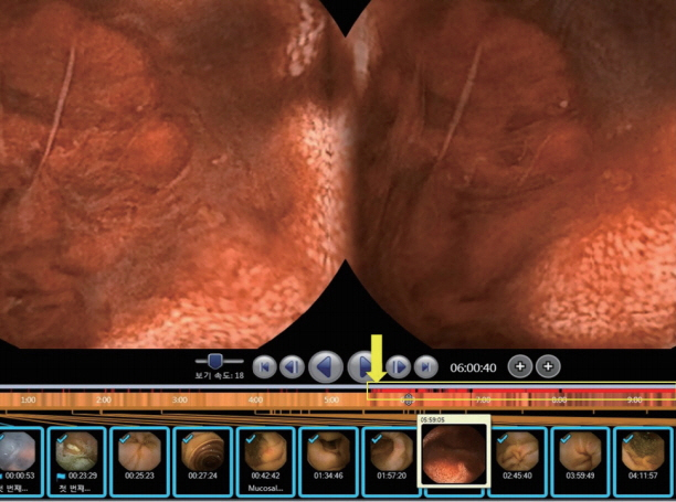
Fig. 1. A representative case with suspected blood indicator showed true blood in the small intestine. Suspected blood indicators are marked with red bands (in the yellow box indicated by a yellow arrow).

Fig. 2. External real-time image viewer.

Fig. 3. External real-time image viewer indicating active small bowel bleeding.
Cited by 1 articles
-
A New Active Locomotion Capsule Endoscopy under Magnetic Control and Automated Reading Program
Dong Jun Oh, Kwang Seop Kim, Yun Jeong Lim
Clin Endosc. 2020;53(4):395-401. doi: 10.5946/ce.2020.127.
Reference
-
1. Iddan G, Meron G, Glukhovsky A, Swain P. Wireless capsule endoscopy. Nature. 2000; 405:417.
Article2. Eliakim R. Video capsule endoscopy of the small bowel. Curr Opin Gastroenterol. 2013; 29:133–139.
Article3. McAlindon ME, Ching HL, Yung D, Sidhu R, Koulaouzidis A. Capsule endoscopy of the small bowel. Ann Transl Med. 2016; 4:369.
Article4. Saurin JC, Beneche N, Chambon C, Pioche M. Challenges and future of wireless capsule endoscopy. Clin Endosc. 2016; 49:26–29.
Article5. Koulaouzidis A, Iakovidis DK, Karargyris A, Plevris JN. Optimizing lesion detection in small-bowel capsule endoscopy: from present problems to future solutions. Expert Rev Gastroenterol Hepatol. 2015; 9:217–235.
Article6. Fisher LR, Hasler WL. New vision in video capsule endoscopy: current status and future directions. Nat Rev Gastroenterol Hepatol. 2012; 9:392–405.
Article7. Goenka MK, Majumder S, Goenka U. Capsule endoscopy: present status and future expectation. World J Gastroenterol. 2014; 20:10024–10037.
Article8. Singeap AM, Stanciu C, Trifan A. Capsule endoscopy: the road ahead. World J Gastroenterol. 2016; 22:369–378.
Article9. Barkin JA, Barkin JS. Video capsule endoscopy: technology, reading, and troubleshooting. Gastrointest Endosc Clin N Am. 2017; 27:15–27.10. Triantafyllou K, Papanikolaou IS, Papaxoinis K, Ladas SD. Two cameras detect more lesions in the small-bowel than one. World J Gastroenterol. 2011; 17:1462–1467.
Article11. Drew K, McAlindon ME, Sanders DS, Sidhu R. The nurse endoscopist: moving ahead with time. Gastroenterol Nurs. 2013; 36:209–213.12. Hakim FA, Alexander JA, Huprich JE, Grover M, Enders FT. CT-enterography may identify small bowel tumors not detected by capsule endoscopy: eight years experience at Mayo Clinic Rochester. Dig Dis Sci. 2011; 56:2914–2919.
Article13. Postgate A, Despott E, Burling D, et al. Significant small-bowel lesions detected by alternative diagnostic modalities after negative capsule endoscopy. Gastrointest Endosc. 2008; 68:1209–1214.
Article14. Zheng Y, Hawkins L, Wolff J, Goloubeva O, Goldberg E. Detection of lesions during capsule endoscopy: physician performance is disappointing. Am J Gastroenterol. 2012; 107:554–560.
Article15. Pezzoli A, Cannizzaro R, Pennazio M, et al. Interobserver agreement in describing video capsule endoscopy findings: a multicentre prospective study. Dig Liver Dis. 2011; 43:126–131.
Article16. Shim KN, Jeon SR, Jang HJ, et al. Quality indicators for small bowel capsule endoscopy. Clin Endosc. 2017; 50:148–160.
Article17. Rajan E, Iyer PG, Oxentenko AS, et al. Training in small-bowel capsule endoscopy: assessing and defining competency. Gastrointest Endosc. 2013; 78:617–622.
Article18. Rondonotti E, Soncini M, Girelli CM, et al. Can we improve the detection rate and interobserver agreement in capsule endoscopy? Dig Liver Dis. 2012; 44:1006–1011.
Article19. Sidhu R, Sakellariou P, McAlindon ME, et al. Is formal training necessary for capsule endoscopy? The largest gastroenterology trainee study with controls. Dig Liver Dis. 2008; 40:298–302.20. Niv Y, Niv G. Capsule endoscopy examination--preliminary review by a nurse. Dig Dis Sci. 2005; 50:2121–2124.
Article21. Dokoutsidou H, Karagiannis S, Giannakoulopoulou E, et al. A study comparing an endoscopy nurse and an endoscopy physician in capsule endoscopy interpretation. Eur J Gastroenterol Hepatol. 2011; 23:166–170.
Article22. Lepileur L, Dray X, Antonietti M, et al. Factors associated with diagnosis of obscure gastrointestinal bleeding by video capsule enteroscopy. Clin Gastroenterol Hepatol. 2012; 10:1376–1380.
Article23. Tacheci I, Devière J, Kopacova M, Douda T, Bures J, Van Gossum A. The importance of upper gastrointestinal lesions detected with capsule endoscopy in patients with obscure digestive bleeding. Acta Gastroenterol Belg. 2011; 74:395–399.24. Buscaglia JM, Giday SA, Kantsevoy SV, et al. Performance characteristics of the suspected blood indicator feature in capsule endoscopy according to indication for study. Clin Gastroenterol Hepatol. 2008; 6:298–301.
Article25. D’Halluin PN, Delvaux M, Lapalus MG, et al. Does the “Suspected Blood Indicator” improve the detection of bleeding lesions by capsule endoscopy? Gastrointest Endosc. 2005; 61:243–249.26. Saurin JC, Lapalus MG, Cholet F, et al. Can we shorten the small-bowel capsule reading time with the “Quick-view” image detection system? Dig Liver Dis. 2012; 44:477–481.
Article27. Westerhof J, Koornstra JJ, Weersma RK. Can we reduce capsule endoscopy reading times? Gastrointest Endosc. 2009; 69:497–502.
Article28. Halling ML, Nathan T, Kjeldsen J, Jensen MD. High sensitivity of quick view capsule endoscopy for detection of small bowel Crohn’s disease. J Gastroenterol Hepatol. 2014; 29:992–996.
Article29. Iakovidis DK, Koulaouzidis A. Software for enhanced video capsule endoscopy: challenges for essential progress. Nat Rev Gastroenterol Hepatol. 2015; 12:172–186.
Article30. Ting DSW, Cheung CY, Lim G, et al. Development and validation of a deep learning system for diabetic retinopathy and related eye diseases using retinal images from multiethnic populations with diabetes. JAMA. 2017; 318:2211–2223.
Article31. Esteva A, Kuprel B, Novoa RA, et al. Dermatologist-level classification of skin cancer with deep neural networks. Nature. 2017; 542:115–118.
Article32. Iakovidis DK, Koulaouzidis A. Automatic lesion detection in capsule endoscopy based on color saliency: closer to an essential adjunct for reviewing software. Gastrointest Endosc. 2014; 80:877–883.
Article33. Karargyris A, Bourbakis N. Detection of small bowel polyps and ulcers in wireless capsule endoscopy videos. IEEE Trans Biomed Eng. 2011; 58:2777–2786.
Article34. Li B, Meng MQ, Lau JY. Computer-aided small bowel tumor detection for capsule endoscopy. Artif Intell Med. 2011; 52:11–16.
Article35. Li BP, Meng MQ. Comparison of several texture features for tumor detection in CE images. J Med Syst. 2012; 36:2463–2469.
Article36. Wu X, Chen H, Gan T, Chen J, Ngo CW, Peng Q. Automatic hookworm detection in wireless capsule endoscopy images. IEEE Trans Med Imaging. 2016; 35:1741–1752.
Article37. Seguí S, Drozdzal M, Vilariño F, et al. Categorization and segmentation of intestinal content frames for wireless capsule endoscopy. IEEE Trans Inf Technol Biomed. 2012; 16:1341–1352.38. Fan Y, Meng MQ, Li B. A novel method for informative frame selection in wireless capsule endoscopy video. Conf Proc IEEE Eng Med Biol Soc. 2011; 2011:4864–4867.39. Rondonotti E, Spada C, Adler S, et al. Small-bowel capsule endoscopy and device-assisted enteroscopy for diagnosis and treatment of small-bowel disorders: European Society of Gastrointestinal Endoscopy (ESGE) technical review. Endoscopy. 2018; 50:423–446.
Article40. Shiotani A, Honda K, Kawakami M, et al. Use of an external real-time image viewer coupled with prespecified actions enhanced the complete examinations for capsule endoscopy. J Gastroenterol Hepatol. 2011; 26:1270–1274.
Article41. Rubin M, Hussain SA, Shalomov A, Cortes RA, Smith MS, Kim SH. Live view video capsule endoscopy enables risk stratification of patients with acute upper GI bleeding in the emergency room: a pilot study. Dig Dis Sci. 2011; 56:786–791.
Article42. Fischer D, Schreiber R, Levi D, Eliakim R. Capsule endoscopy: the localization system. Gastrointest Endosc Clin N Am. 2004; 14:25–31.
Article43. Slawinski PR, Obstein KL, Valdastri P. Capsule endoscopy of the future: what’s on the horizon? World J Gastroenterol. 2015; 21:10528–10541.
Article44. Marya N, Karellas A, Foley A, Roychowdhury A, Cave D. Computerized 3-dimensional localization of a video capsule in the abdominal cavity: validation by digital radiography. Gastrointest Endosc. 2014; 79:669–674.
Article45. Slawinski PR, Obstein KL, Valdastri P. Emerging issues and future developments in capsule endoscopy. Tech Gastrointest Endosc. 2015; 17:40–46.
Article46. Li X, Chen H, Dai J, Gao Y, Ge Z. Predictive role of capsule endoscopy on the insertion route of double-balloon enteroscopy. Endoscopy. 2009; 41:762–766.
Article47. Karargyris A, Koulaouzidis A. OdoCapsule: next-generation wireless capsule endoscopy with accurate lesion localization and video stabilization capabilities. IEEE Trans Biomed Eng. 2015; 62:352–360.
Article
- Full Text Links
-
- Actions
-
Cited
- CITED
-
- Close
- Share
-
- Similar articles
-
- Small Bowel Obstruction and Capsule Retention by a Small Bowel Ulcer That Was Not Found on Capsule Endoscopy
- Capsule Endoscopy in Children
- The Future of Capsule Endoscopy: The Role of Artificial Intelligence and Other Technical Advancements
- The Role of Capsule Endoscopy in the Diagnosis of Crohn's Disease
- Current status and future perspectives of capsule endoscopy
