Profiling of Serum Metabolites Using MALDI-TOF and Triple-TOF Mass Spectrometry to Develop a Screen for Ovarian Cancer
- Affiliations
-
- 1Biomarker Branch, Research Institute, National Cancer Center, Goyang, Korea. yoo_akh@ncc.re.kr
- 2Division of Gynecologic Oncology, Department of Obstetrics and Gynecology, Ewha Womans University Mokdong Hospital, Ewha Womans University School of Medicine, Seoul, Korea. yoo_akh@ncc.re.kr
- 3Department of Genetic Engineering, Sungkyunkwan University, Suwon, Korea.
- KMID: 2417877
- DOI: http://doi.org/10.4143/crt.2017.275
Abstract
- PURPOSE
We sought to develop a matrix assisted laser desorption ionization-time of flight (MALDI-TOF)-based, ovarian cancer (OVC), low-mass-ion discriminant equation (LOME) and to evaluate a possible supportive role for triple-TOF mass analysis in identifying metabolic biomarkers.
MATERIALS AND METHODS
A total of 114 serum samples from patients with OVC and benign ovarian tumors were subjected to MALDI-TOF analysis and a total of 137 serum samples from healthy female individuals and patients with OVC, colorectal cancer, hepatobiliary cancer, and pancreatic cancer were subjected to triple-TOF analysis. An OVC LOME was constructed by reference to the peak intensity ratios of discriminatory low-mass ion (LMI) pairs. Triple-TOF analysiswas used to select and identify metabolic biomarkers for OVC screening.
RESULTS
Three OVC LOMEs were finally constructed using discriminatory LMI pairs (137.1690 and 84.4119 m/z; 496.5022 and 709.7642 m/z; and 524.5614 and 709.7642 m/z); all afforded accuracies of > 90%. The LMIs at 496.5022 m/z and 524.5614 m/z were those of lysophosphatidylcholine (LPC) 16:0 and LPC 18:0. Triple-TOF analysis selected seven discriminative LMIs; each LMI had a specificity > 90%. Of the seven LMIs, fourwith a 137.0455 m/z ion atretention times of 2.04-3.14 minuteswere upregulated in sera from OVC patients; the ion was identified as that derived from hypoxanthine.
CONCLUSION
MALDI-TOF-based OVC LOMEs combined with triple-TOF-based OVC metabolic biomarkers allow reliable OVC screening; the techniques are mutually complementary both quantitatively and qualitatively.
MeSH Terms
Figure
-

Fig. 1. Separation results obtained during one of the six repeats of matrix assisted laser desorption ionization-time of flight analysis. (A) Principal component analysis-based discriminant analysis (PCA-DA). (B) Preliminary low-mass-ion candidates. DS, discriminant score; OVC, ovarian cancer; BOT, benign ovarian tumor.
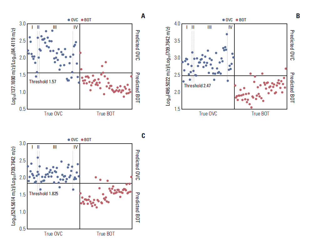
Fig. 2. Discriminatory results afforded by the ratios of low-mass ion pairs on matrix assisted laser desorption ionization-time of flight analysis. (A) Log10(137.1690 m/z)/Log10(84.4119 m/z). (B) Log10(496.5022 m/z)/Log10(709.7642 m/z). (C) Log10 (524.5614 m/z)/Log10(709.7642 m/z). OVC, ovarian cancer; BOT, benign ovarian tumor.
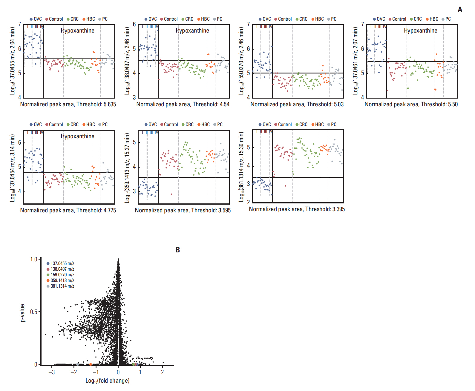
Fig. 3. Seven candidate metabolites suggested by triple-TOF analysis to be characteristic of ovarian cancer (OVC). (A) Figure shows relative amount of each candidate metabolites [Y-axis, Log10(mass peak area of low-mass ion)] in healthy women (control) and those with OVC, colorectal cancer (CRC), hepatobiliary cancer (HBC), and pancreatic cancer (PC). Low-mass ions (LMIs) of 137.0455 m/z, retention time (RT) 2.04 min; 138.0497 m/z, RT 2.46 min; 159.0270 m/z, RT 2.46 min; 137.0461 m/z, RT 2.07 min; 137.0454 m/z, RT 3.14 min; 359.1413 m/z, RT 15.27 min; 381.1314 m/z, RT 15.30 min. Of the seven LMIs, four with a 137.0455 m/z ion at RTs of 2.04-3.14 min were upregulated in sera from OVC patients; those LMIs were identified as hypoxanthine. (B) A volcano plot of low-mass ions detected on triple-TOF analysis.
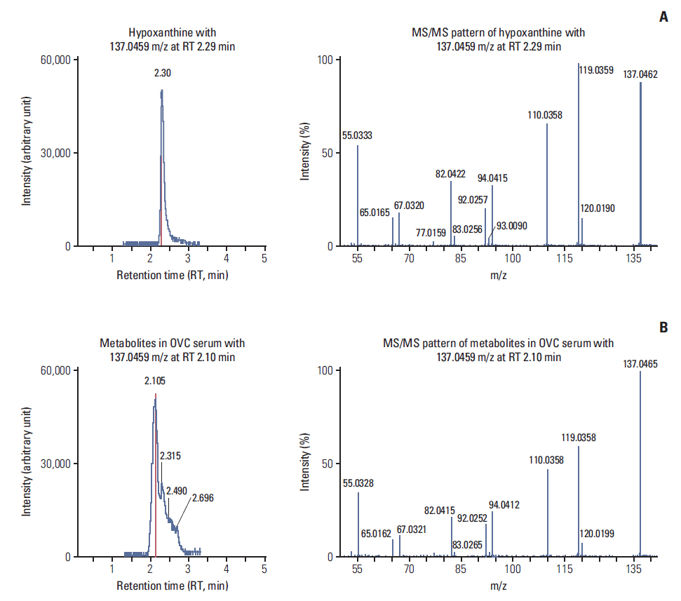
Fig. 4. Identification of hypoxanthine upregulated in sera of ovarian cancer (OVC) patients. A low-mass ion of 137.0455 m/z (left panel, B) exhibited a retention time (RT) that differed slightly from that of hypoxanthine (left panel, A), but the tandem mass spectrometry (MS/MS) pattern (right panel, B) was exactly that of hypoxanthine (right panel, A).
Reference
-
References
1. Kim YH, Kim SC. Recent advances in the biomarkers for epithelial ovarian cancer. J Gynecol Oncol. 2011; 22:219–21.
Article2. Menon U, Gentry-Maharaj A, Hallett R, Ryan A, Burnell M, Sharma A, et al. Sensitivity and specificity of multimodal and ultrasound screening for ovarian cancer, and stage distribution of detected cancers: results of the prevalence screen of the UK Collaborative Trial of Ovarian Cancer Screening (UKCTOCS). Lancet Oncol. 2009; 10:327–40.3. Jacobs IJ, Menon U, Ryan A, Gentry-Maharaj A, Burnell M, Kalsi JK, et al. Ovarian cancer screening and mortality in the UK Collaborative Trial of Ovarian Cancer Screening (UKCTOCS): a randomised controlled trial. Lancet. 2016; 387:945–56.4. Buys SS, Partridge E, Black A, Johnson CC, Lamerato L, Isaacs C, et al. Effect of screening on ovarian cancer mortality: the Prostate, Lung, Colorectal and Ovarian (PLCO) Cancer Screening Randomized Controlled Trial. JAMA. 2011; 305:2295–303.5. Lee JH, Yoo BC, Kim YH, Ahn SA, Yeo SG, Cho JY, et al. Lowmass-ion discriminant equation (LOME) for ovarian cancer screening. BioData Min. 2016; 9:32.
Article6. Lee JH, Kim KH, Park JW, Chang HJ, Kim BC, Kim SY, et al. Low-mass-ion discriminant equation: a new concept for colorectal cancer screening. Int J Cancer. 2014; 134:1844–53.
Article7. Kim K, Yeo SG, Yoo BC. Identification of hypoxanthine and phosphoenolpyruvic acid as serum markers of chemoradiotherapy response in locally advanced rectal cancer. Cancer Res Treat. 2015; 47:78–89.
Article8. Yoo BC, Kong SY, Jang SG, Kim KH, Ahn SA, Park WS, et al. Identification of hypoxanthine as a urine marker for non-Hodgkin lymphoma by low-mass-ion profiling. BMC Cancer. 2010; 10:55.
Article9. Lorenzi M, Vannoni D, Leoncini R, Caldarone R, Marinello E. The determination of urinary oxypurines as markers of gastrointestinal tumors. Tumori. 1987; 73:289–94.
Article10. Jung J, Jung Y, Bang EJ, Cho SI, Jang YJ, Kwak JM, et al. Noninvasive diagnosis and evaluation of curative surgery for gastric cancer by using NMR-based metabolomic profiling. Ann Surg Oncol. 2014; 21 Suppl 4:S736–42.
Article11. Kyriakides M, Rama N, Sidhu J, Gabra H, Keun HC, El-Bahrawy M. Metabonomic analysis of ovarian tumour cyst fluid by proton nuclear magnetic resonance spectroscopy. Oncotarget. 2016; 7:7216–26.
Article12. Chan TC, Howell SB. Role of hypoxanthine and thymidine in determining methotrexate plus dipyridamole cytotoxicity. Eur J Cancer. 1990; 26:907–11.
Article
- Full Text Links
-
- Actions
-
Cited
- CITED
-
- Close
- Share
-
- Similar articles
-
- Taxonomic Identification of Bacillus Species Using Matrix-Assisted Laser Desorption/Ionization-Time of Flight Mass Spectrometry
- Identification of biomarker for ovarian cancer by serum proteomic analysis using SELDI-ToF-MS
- Matrix-Assisted Laser Desorption Ionization Time-of-Flight Mass Spectrometry in Clinical Microbiology: What Are the Current Issues?
- Evaluation of Matrix-Assisted Laser Desorption Ionization-Time of Flight Mass Spectrometry for Identification of Aerobic Bacteria in a Clinical Microbiology Laboratory
- Reliability of Acinetobacter baumannii Identification with Matrix-Assisted Laser Desorption Ionization-Time of Flight Mass Spectrometry
