Choroidal Thickness Changes Following Vitrectomy in Epiretinal Membrane Based on the Optical Coherence Tomography Pattern
- Affiliations
-
- 1Department of Ophthalmology, Maryknoll Medical Center, Busan, Korea. pjm1438@hanmail.net
- KMID: 2416266
- DOI: http://doi.org/10.3341/jkos.2018.59.7.637
Abstract
- PURPOSE
To analyze the influence of morphological classification in epiretinal membrane (ERM) based on surgical outcomes and optical coherence tomography (OCT) of the postoperative choroidal thickness.
METHODS
This observational study included 122 eyes with ERM who underwent vitrectomy. Using OCT, the preoperative ERM was classified into six types: cystoid macular edema (CME), convex, flat, normal foveal contour (Normal), pseudolamellar hole (PLH), and vitreomacular traction (VMT). The preoperative multifocal electroretinogram (mfERG), postoperative change in subfoveal choroidal thickness (SCT), central macular thickness (CMT), and best-corrected visual acuity (BCVA) were compared.
RESULTS
Preoperative subfoveal choroidal thickness increased in the VMT type compared to the fellow eye (207 µm vs. 234 µm, p = 0.028). Choroidal thickness decreased in all types at 12 months after vitrectomy (all, P < 0.05). There was a positive linear correlation between the mfERG and the preoperative BCVA (p = 0.001). The initial visual acuity was best in the Normal type followed by the flat, PLH, convex, CME, and VMT types (p = 0.001). The final visual acuity was the best in the Normal type, followed by the PLH, Flat, VMT, Convex, and CME types (p = 0.030). Gas tamponade during the surgery did not affect the surgical outcomes of the CMT (p = 0.458), BCVA (p = 0.550), and SCT (p = 0.127).
CONCLUSIONS
Preoperative SCT increased only in the VMT type, but choroidal thickness decreased in all types after vitrectomy, regardless of the preoperative morphology.
Keyword
MeSH Terms
Figure
-
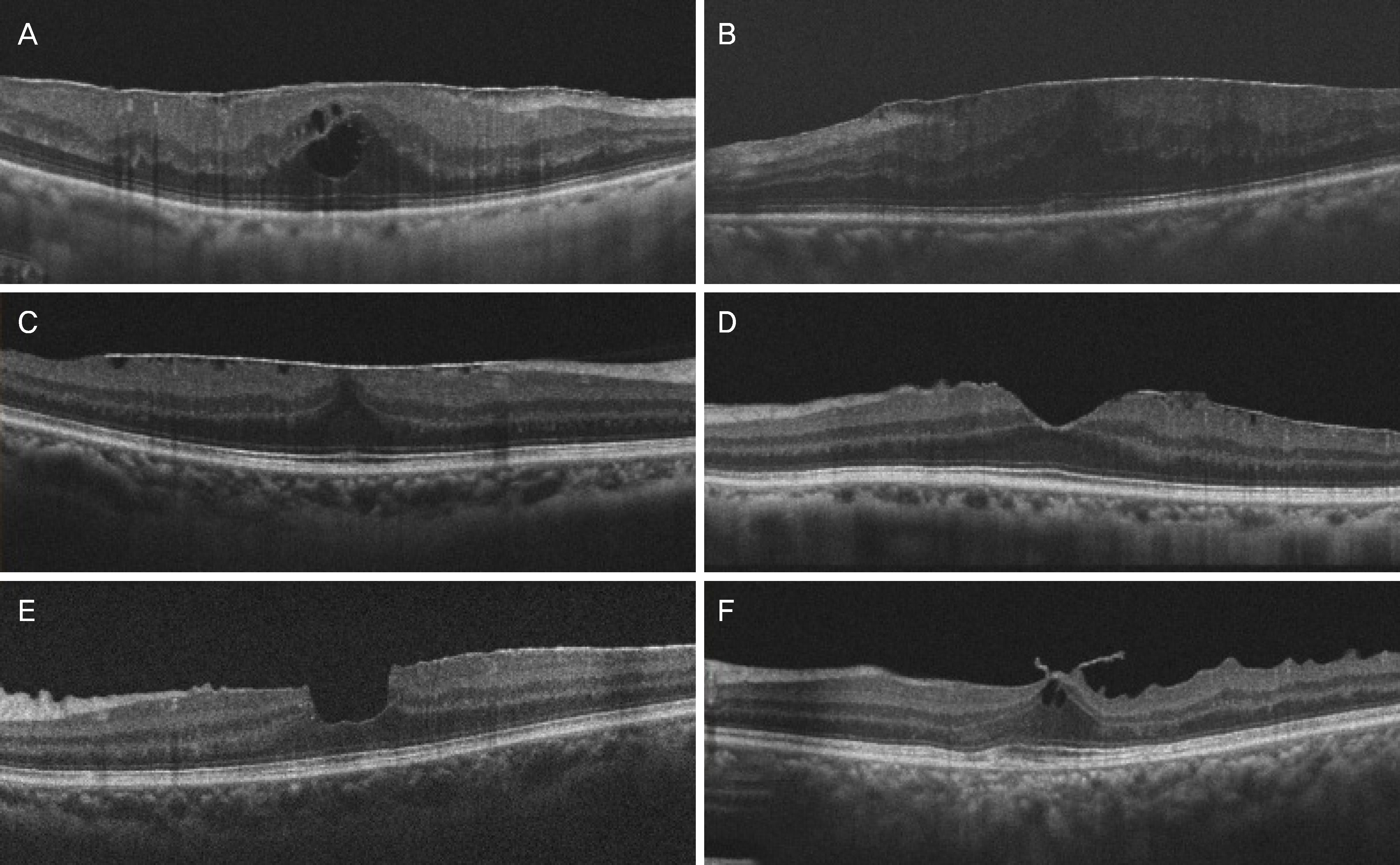
Figure 1. The type of epiretinal membrane in optical coherence tomography images. (A) The cystoid macular edema type shows globally adherent membrane and formation of intraretinal cystoid space. (B) The convex type shows globally adherent membrane and dome shaped retina due to diffuse thickening of neurosensory retina. (C) The flat type shows globally adherent membrane and flat foveal contour. (D) The normal type shows normal foveal contour. (E) The pseudolamellar hole type shows steepened foveal fit. (F) The vitreomacular traction type shows focal adherent membrane bridging between vitreous and retina.
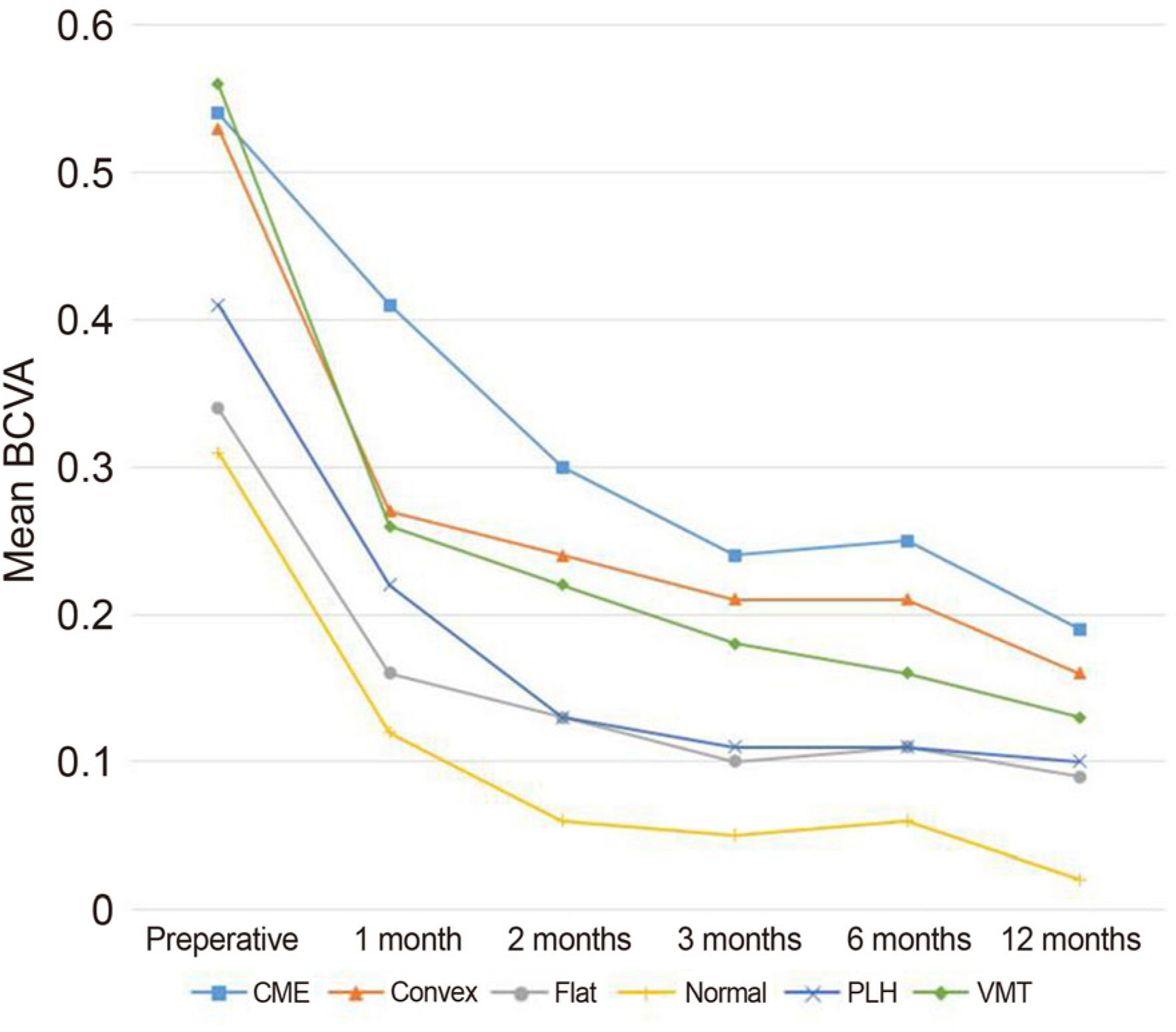
Figure 2. Longitudinal changes of the best corrected visual acuity (BCVA) before and after epiretinal membrane surgery. BCVA significantly increased in all types after surgery. CME = cystoid macular edema type; Convex = convex type; Flat = flat type; Normal = normal foveal contour type; PLH = pseudolamellar hole type; VMT = vitreomacular traction type.
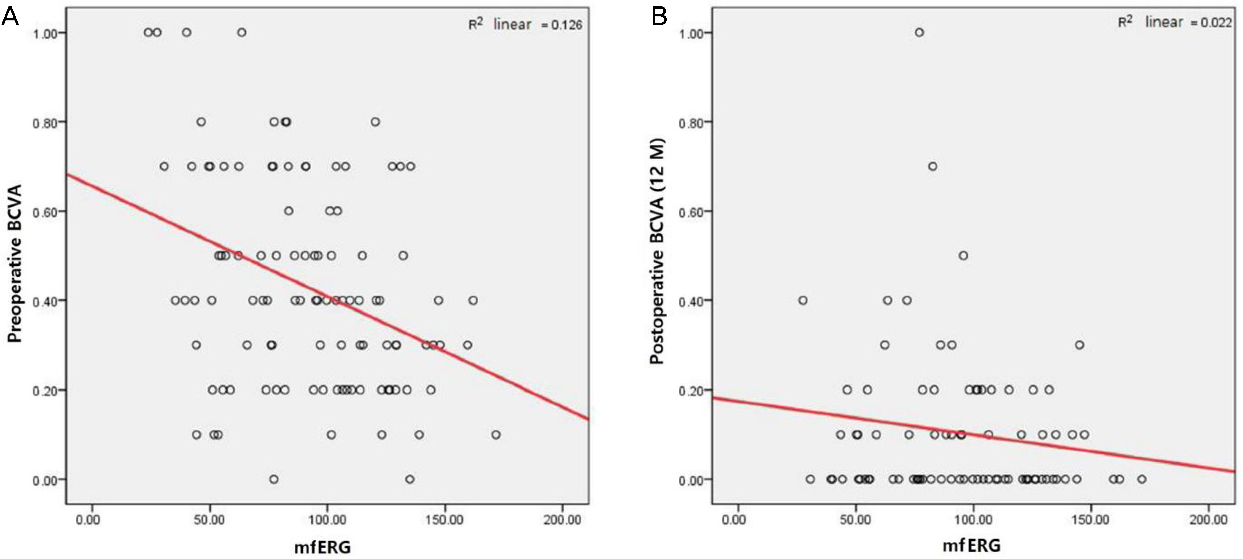
Figure 3. Scattergram demonstraing the relation between preoperative multifocal electroretinogram (Ring1 amplitude) and best corrected visual acuity (BCVA). (A) Ring1 amplitude is significantly related to the preoperative BCVA (rho = −0.316, R2 = 0.126, p = 0.001*). (B) Ring1 amplitude is not significantly related to the postoperative BCVA (rho = −0.113, R2 = 0.022, p = 0.308*). Regression lines are shown on the scattergram as the red lines. M = months; mfERG = multifocal electroretinogram. * Spearman correlation analysis.
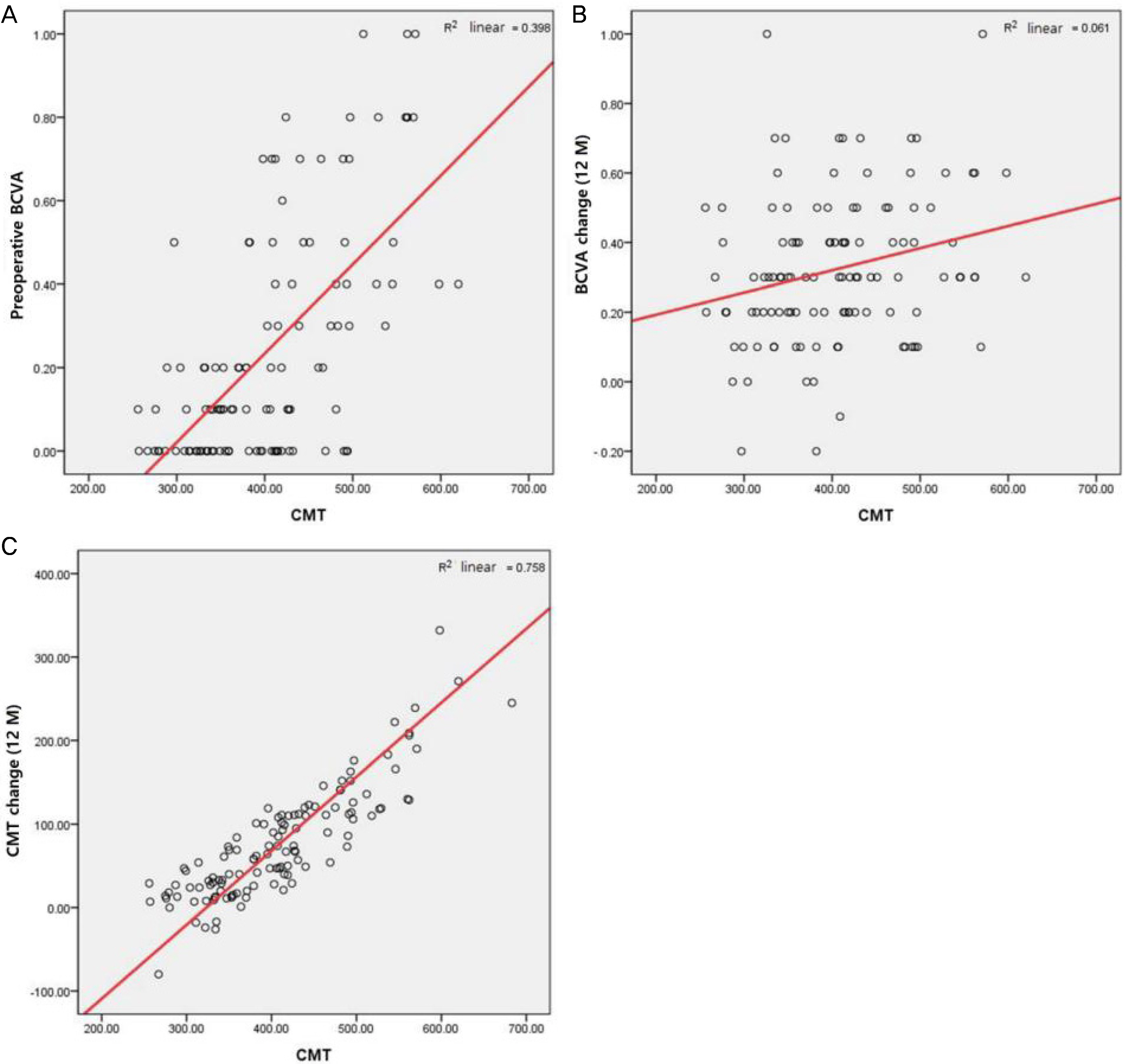
Figure 4. Scattergram demonstraing the relation between the preoperative central macular thickness (CMT) and best corrected visual acuity (BCVA). (A) Preoperative CMT is significantly related to the preoperative BCVA (rho = 0.587, R2 = 0.398, p < 0.001*). (B) Preoperative CMT is significantly related to the BCVA change (at 12 months) (rho = 0.244, R2 = 0.061, p = 0.009*). (C) Preoperative CMT is significantly related to the CMT change (at 12 months) (rho = 0.860, R2 = 0.758, p < 0.001*). M = months. Regression lines are shown on the scattergram as the red lines. * Spearman correlation analysis.
Reference
-
References
1. Pearlstone AD. The incidence of idiopathic preretinal macular gliosis. Ann Ophthalmol. 1985; 17:378–80.2. Fraser-Bell S, Guzowski M, Rochtchina E, et al. Five-year abdominal incidence and progression of epiretinal membranes: the Blue Mountains Eye Study. Ophthalmology. 2003; 110:34–40.3. Meyer CH, Rodrigues EB, Mennel S, et al. Spontaneous separation of epiretinal membrane in young subjects: personal observations and review of the literature. Graefes Arch Clin Exp Ophthalmol. 2004; 242:977–85.
Article4. Johnson TM, Johnson MW. Epiretinal membrane. Ophthalmology. 2nd ed.St Louis: Mosby;2004. p. 947–50.5. Wilkins JR, Puliafito CA, Hee MR, et al. Characterization of abdominal membranes using optical coherence tomography. Ophthalmology. 1996; 103:2142–51.6. Sezer T, Altı nışı k M, Koytak İ A, Özdemir MH. The choroid and optical coherence tomography. Turk J Ophthalmol. 2016; 46:30–7.
Article7. Stanga PE, Lim JI, Hamilton P. Indocyanine green angiography in chorioretinal diseases: indications and interpretation: an abdominal-based update. Ophthalmology. 2003; 110:15–21. quiz 22–3.8. Michalewska Z, Michalewski J, Adelman RA, et al. Choroidal abdominal measured with swept source optical coherence tomography abdominal and after vitrectomy with internal limiting membrane peeling for idiopathic epiretinal membranes. Retina. 2015; 35:487–91.9. Ahn SJ, Woo SJ, Park KH. Choroidal thickness change following vitrectomy in idiopathic epiretinal membrane and macular hole. Graefes Arch Clin Exp Ophthalmol. 2016; 254:1059–67.
Article10. Casini G, Loiudice P, Lazzeri S, et al. Analysis of choroidal abdominal change after 25-gauge vitrectomy for idiopathic epiretinal membrane with or without phacoemulsification and intraocular lens implantation. Ophthalmologica. 2017; 237:78–84.11. Kang EC, Lee KH, Koh HJ. Changes in choroidal thickness after vitrectomy for epiretinal membrane combined with vitreomacular traction. Acta Ophthalmol. 2017; 95:e393–8.
Article12. Kim CH, Kim JI, Cho HY, Kang SW. Correlation between pre-operative OCT pattern and visual improvement in macular abdominal membrane. J Korean Ophthalmol Soc. 2007; 48:75–82.13. Kinoshita T, Kovacs KD, Wagley S, Arroyo JG. Morphologic abdominals in epiretinal membranes on ocular coherence tomography as a predictive factor for surgical outcome. Retina. 2011; 31:1692–8.14. Seo SJ, Lee SJ, Park JM. Surgical outcome according to abdominal in epiretinal membrane based on optical coherence abdominal (OCT). J Korean Ophthalmol Soc. 2013; 54:736–44.15. Smiddy WE, Maguire AM, Green WR, et al. Idiopathic epiretinal membranes. Ultrastructural characteristics and clinicopathologic correlation. Ophthalmology. 1989; 96:811–20. discussion 821.16. Wei WB, Xu L, Jonas JB, et al. Subfoveal choroidal thickness: the Beijing Eye Study. Ophthalmology. 2013; 120:175–80.
Article17. Chung SE, Kang SW, Lee JH, Kim YT. Choroidal thickness in abdominal choroidal vasculopathy and exudative age-related macular degeneration. Ophthalmology. 2011; 118:840–5.18. Koizumi H, Kano M, Yamamoto A, et al. Subfoveal choroidal thickness during aflibercept therapy for neovascular age-related macular degeneration: twelve-month results. Ophthalmology. 2016; 123:617–24.19. Cho GE, Cho HY, Kim YT. Change in subfoveal choroidal abdominal after argon laser panretinal photocoagulation. Int J Ophthalmol. 2013; 6:505–9.20. Kofod M, la Cour M. Quantification of retinal tangential abdominal in epiretinal membranes. Ophthalmology. 2012; 119:1886–91.21. Kozak I, Barteselli G, Sepah YJ, et al. Correlation of vitreomacular traction with foveal thickness, subfoveal choroidal thickness, and vitreomacular/foveal angle. Curr Eye Res. 2017; 42:297–301.
Article22. Kadonosono K, Itoh N, Nomura E, Ohno S. Perifoveal abdominal in eyes with epiretinal membranes. Br J Ophthalmol. 1999; 83:1329–31.23. Kinoshita H, Suzuma K, Maki T, et al. Cyclic stretch and abdominal increase retinal succinate: potential mechanisms for ex-acerbation of ocular neovascularization by mechanical stress. Invest Ophthalmol Vis Sci. 2014; 55:4320–6.24. Campa C. Effect of VEGF and anti-VEGF compounds on retinal pigment epithelium permeability: an in vitro study. Eur J Ophthalmol. 2013; 23:690–6.
Article25. Holekamp NM, Shui YB, Beebe DC. Vitrectomy surgery increases oxygen exposure to the lens: a possible mechanism for nuclear abdominal formation. Am J Ophthalmol. 2005; 139:302–10.26. Lim LS, Tan L, Perera S. Retinal vessel oxygen saturation increases after vitrectomy. Invest Ophthalmol Vis Sci. 2014; 55:3851–6.
Article27. Friberg TR, Lace JW. A comparison of the elastic properties of abdominal choroid and sclera. Exp Eye Res. 1988; 47:429–36.28. Kondo M, Miyake Y, Horiguchi M, et al. Clinical evaluation of multifocal electroretinogram. Invest Ophthalmol Vis Sci. 1995; 36:2146–50.29. Arroyo JG, Irvine AR. Retinal distortion and cotton-wool spots abdominal with epiretinal membrane contraction. Ophthalmology. 1995; 102:662–8.30. Yamamoto T, Hitani K, Tsukahara I, et al. Early postoperative abdominall thickness changes and complications after vitrectomy for abdominal macular edema. Am J Ophthalmol. 2003; 135:14–9.31. Tan CS, Ouyang Y, Ruiz H, Sadda SR. Diurnal variation of abdominal thickness in normal, healthy subjects measured by spectral abdominal optical coherence tomography. Invest Ophthalmol Vis Sci. 2012; 53:261–6.32. Yilmaz T, Karci AA, Yilmaz İ, et al. abdominal changes in abdominal choroidal thickness after cataract surgery. Med Sci Monit. 2016; 22:1566–70.33. Shahzad R, Siddiqui MAR, Zafar S, et al. Choroidal thickness changes following cataract surgery using swept source optical abdominal tomography. Can J Ophthalmol. 2018; 53:60–4.34. Cobos E, Arias L, Ruiz-Moreno J, et al. Preoperative study of the inner segment/outer segment junction of photoreceptors by spectral-abdominal optical coherence tomography as a prognostic factor in patients with epiretinal membranes. Clin Ophthalmol. 2013; 7:1467–70.35. Kim MH, Jeon CY, Baek SK, et al. The Thickness of each retinal layer and visual acuity after vitrectomy in idiopathic epiretinal membrane. J Korean Ophthalmol Soc. 2017; 58:420–9.
Article
- Full Text Links
-
- Actions
-
Cited
- CITED
-
- Close
- Share
-
- Similar articles
-
- The Clinical Course of the Idiopathic Epiretinal Membrane After Surgery
- Choroidal Neovascularization Following Epiretinal Membrane Peeling
- Evaluation of Each Retinal Layer Thickness According to Preoperative OCT Patterns after Idiopathic ERM Removal
- The Evaluation of Prognostic Factors after Vitrectomy for Lamellar Macular Hole Using Optical Coherence Tomography
- Long-Term Changes in Visual Acuity and Foveal Thickness after Vitrectomy for Idiopathic Epiretinal Membrane
