Korean J Ophthalmol.
2015 Dec;29(6):439-440. 10.3341/kjo.2015.29.6.439.
Choroidal Neovascularization Following Epiretinal Membrane Peeling
- Affiliations
-
- 1Department of Ophthalmology, Seoul National University Bundang Hospital, Seoul National University College of Medicine, Seongnam, Korea. sejoon1@snu.ac.kr
- KMID: 2363855
- DOI: http://doi.org/10.3341/kjo.2015.29.6.439
Abstract
- No abstract available.
MeSH Terms
Figure
-
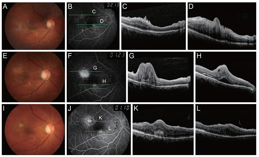
Fig. 1 Fundus photograph of the right eye showed drusenoid deposit and macular pucker (A). Fundus fluorescein angiography revealed multiple hyperfluorescent lesions around the epiretinal membrane (ERM) (B). Optical coherence tomography showed ERM with macular edema in the right eye (C,D). Postoperative fundus photograph revealed satisfactory ERM removal (E). Fluorescein angiography showed leakage superotemporal to the fovea in the early phase (F), and optical coherence tomography revealed extrafoveal choroidal neovascularization combined with subretinal fluid (G,H). After several intravitreal aflibercept injections, choroidal neovascularization regressed and no subretinal fluid was observed (I-L).
Reference
-
1. Fang X, Chen Z, Weng Y, et al. Surgical outcome after removal of idiopathic macular epiretinal membrane in young patients. Eye (Lond). 2008; 22:1430–1435.2. Goh YW, Ehrlich R, Welch S. Iatrogenic choroidal neovascularization following idiopathic epiretinal membrane peel. Digit J Ophthalmol. 2013; 19:9–12.3. Warden SM, Pachydaki SI, Christoforidis JB, et al. Choroidal neovascularization after epiretinal membrane removal. Arch Ophthalmol. 2006; 124:1652–1654.
- Full Text Links
-
- Actions
-
Cited
- CITED
-
- Close
- Share
-
- Similar articles
-
- Effects of Internal Limiting Membrane Peeling in Combined Hamartoma of Retina and Retinal Pigment Epithelium
- Analysis of Leading Diseases Causing Epiretinal Membrane and Comparison of Prognosis after Epiretinal Membrane Peeling
- The Effect of Internal Limiting Membrane Peeling in Treatment of Idiopathic Epiretinal Membrane
- The Effect of Internal Limiting Membrane Peeling in Epiretinal Membrane Including Pseudolamellar Macular Hole
- Internal Limiting Membrane Peeling In Surgical Treatment of Macular Epiretinal Membrane
