Effects of Internal Limiting Membrane Peeling in Combined Hamartoma of Retina and Retinal Pigment Epithelium
- Affiliations
-
- 1Department of Ophthalmology, Maryknoll Medical Center, Busan, Korea. pjm1438@hanmail.net
- KMID: 2401674
- DOI: http://doi.org/10.3341/jkos.2018.59.1.23
Abstract
- PURPOSE
To evaluate the effects of peeling the internal limiting membrane (ILM) of an epiretinal membrane (ERM) for surgical treatment of combined hamartoma of the retina and retinal pigment epithelium (RPE).
METHODS
We retrospectively reviewed the records of 11 patients (11 eyes) with ERM of combined hamartoma of the retina + RPE and 22 patients (22 eyes) with idiopathic ERM who treated with pars plana vitrectomy and removal of the ERM. The patients were divided into four groups: involving eyes with (5 eyes) or without (6 eyes) ILM peeling in combined hamartoma of the retina + RPE group and eyes with (10 eyes) or without (12 eyes) ILM peeling in the idiopathic ERM group. Anatomical outcomes, functional outcomes, complications and recurrences were compared between eyes with and without ILM peeling.
RESULTS
Central retinal thickness (CRT) and subfoveal choroidal thickness (SFCT) decreased and postoperative best-corrected visual acuity (BCVA) improved regardless of ILM peeling in all groups. There were no statistically significant differences in CRT, SFCT and BCVA between without and with ILM peeling in ERM of combined hamartoma of the retina + RPE and idiopathic ERM groups. There were no complications and recurrences in any group.
CONCLUSIONS
The additional ILM peeling did not affect the postoperative results.
Keyword
MeSH Terms
Figure
-
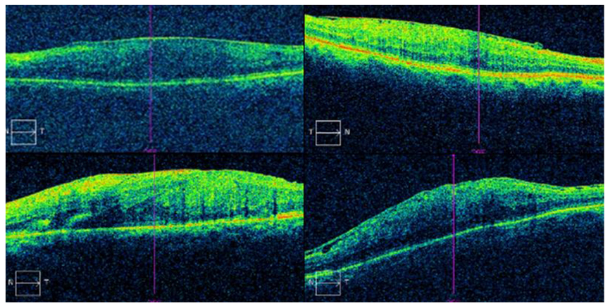
Figure 1 Preoperative optical coherence tomography images of combined hamartoma of the retina and retinal pigment epithelium. These images showed the epiretinal membrane of combined hamartoma of the retina and retinal pigment epithelium and distorted fovea contour.

Figure 2 Epiretinal membrane (ERM) of combined hamartoma of the retina and retinal pigment epithelium in 73 years old man. (A) Preoperative fundus photo showed gray elevated lesion with preretinal gliosis, vascular tortuosity with vascular traction, hyperplasia of retinal pigment epithelium at the margin of growth. Preoperative visual acuity was logMAR 0.5. (B) Preoperative enhanced depth image (EDI) of optical coherence tomography showed folded, distorted retinal thickening with ERM. (C) Preoperative fluorescein angiography showed increasing fluorescence in perimacular area in early phase. (D) Preoperative fluorescein angiography showed hyperfluorescent lesion with blocked fluorescence, vascular distortion with retinal vascular contracting toward the lesion in late phase. (E) Fundus photo at 12 months after membrane peeling, internal limiting membrane peeling, ERM of combined hamartoma of the retina and retinal pigment epithelium was removed. Visual acuity was improved to logMAR 0.1. (F) EDI at 12 months after membrane peeling, internal limiting membrane peeling, central retinal thickness value has decreased 550 µm to 337 µm and subfoveal choroidal thickness value has decreased 203 µm to 191 µm.
Reference
-
1. Shields CL, Thangappan A, Hartzell K, et al. Combined hamartoma of the retina and retinal pigment epithelium in 77 consecutive patients visual outcome based on macular versus extramacular tumor location. Ophthalmology. 2008; 115:2246–2252.e3.2. Shields CL, Mashayekhi A, Dai VV, et al. Optical coherence tomographic findings of combined hamartoma of the retina and retinal pigment epithelium in 11 patients. Arch Ophthalmol. 2005; 123:1746–1750.
Article3. Park DW, Dugel PU, Garda J, et al. Macular pucker removal with and without internal limiting membrane peeling: pilot study. Ophthalmology. 2003; 110:62–64.
Article4. Kwok AK, Lai TY, Li WW, et al. Indocyanine green-assisted internal limiting membrane removal in epiretinal membrane surgery: a clinical and histologic study. Am J Ophthalmol. 2004; 138:194–199.
Article5. Kim TW, Song SJ, Chung H, Yu HG. Internal limiting membrane peeling in surgical treatment of macular epiretinal membrane. J Korean Ophthalmol Soc. 2005; 46:989–994.6. Kim SK, Chung JW, Chung H, Yu HG. Phacoemulsification and foldable intraocular lens implantation combined with vitrectomy and silicone oil tamponade for severe proliferative diabetic retinopathy. J Cataract Refract Surg. 2004; 30:1721–1726.
Article7. Simpson AR, Petrarca R, Jackson TL. Vitreomacular adhesion and neovascular age-related macular degeneration. Surv Ophthalmol. 2012; 57:498–509.
Article8. Shah SU, Haller JA. Vitreomacular traction in a case of exudative age-related macular degeneration resistant to anti-VEGF therapy. Acta Ophthalmol. 2012; 90:e569–e570.
Article9. Kinoshita H, Suzuma K, Maki T, et al. Cyclic stretch and hypertension increase retinal succinate: potential mechanisms for exacerbation of ocular neovascularization by mechanical stress. Invest Ophthalmol Vis Sci. 2014; 55:4320–4326.
Article10. Nickla DL, Wallman J. The multifunctional choroid. Prog Retin Eye Res. 2010; 29:144–168.
Article11. Kang EC, Lee KH, Koh HJ. Changes in choroidal thickness after vitrectomy for epiretinal membrane combined with vitreomacular traction. Acta Ophthalmol. 2017; 95:e393–e398.
Article12. Aung KZ, Makeyeva G, Adams MK, et al. The prevalence and risk factors of epiretinal membranes: the Melbourne Collaborative Cohort Study. Retina. 2013; 33:1026–1034.13. Shea M. The surgical management of macular pucker in rhegmatogenous retinal detachment. Ophthalmology. 1980; 87:70–74.
Article14. Sivalingam A, Eagle RC Jr, Duker JS, et al. Visual prognosis correlated with the presence of internal-limiting membrane in histopathologic specimens obtained from epiretinal membrane surgery. Ophthalmology. 1990; 97:1549–1552.15. Engelbrecht NE, Freeman J, Sternberg P Jr, et al. Retinal pigment epithelial changes after macular hole surgery with indocyanine green-assisted internal limiting membrane peeling. Am J Ophthalmol. 2002; 133:89–94.
Article16. Uemura A, Kanda S, Sakamoto Y, Kita H. Visual field defects after uneventful vitrectomy for epiretinal membrane with indocyanine green-assisted internal limiting membrane peeling. Am J Ophthalmol. 2003; 136:252–257.
Article17. Gass JDM. Stereoscopic Atlas of Macular Disease: Diagnosis and Treatment. 3rd ed. Vol. 2. St. Louis: Mosby;1987. p. 676–693.
- Full Text Links
-
- Actions
-
Cited
- CITED
-
- Close
- Share
-
- Similar articles
-
- Spectral-domain Optical Coherence Tomography of Combined Hamartoma of the Retina and Retinal Pigment Epithelium in Neurofibromatosis
- Epiretinal Membrane of Combined Hamartoma of Retina and Retinal Pigment Epithelium Versus Idiopathic Epiretinal Membrane
- Histologic Study in Transplantation of Cultured Rabbit Retinal Pigment Epithelium
- Effect of Internal Limiting Membrane Peeling in Idiopathic Macular Holes Stage 3, 4
- Tomographic Structural Changes of the Inner Retina after Internal Limiting Membrane Peeling for Idiopathic Epiretinal Membrane
