J Bacteriol Virol.
2018 Jun;48(2):49-58. 10.4167/jbv.2018.48.2.49.
Novel Cell Permeable Peptide Based on Multi Basic Furin-dependent Cleavage Site of Respiratory Syncytial Virus Fusion Protein
- Affiliations
-
- 1Division of Viral Disease Research, Center for Infectious Diseases Research, Korea National Institute of Health, Cheongju, Korea. tigerkis@nih.go.kr
- 2Division of Viral Diseases, Center for Laboratory Control of Infectious Diseases, Korea Centers for Disease Control and Prevention, Cheongju, Korea. chunkang@korea.kr
- 3Department of Microbiology, Chungbuk National University, Cheongju, Korea.
- 4Department of Life Science and Technology, Pai Chai University, Daejeon, Korea.
- KMID: 2414855
- DOI: http://doi.org/10.4167/jbv.2018.48.2.49
Abstract
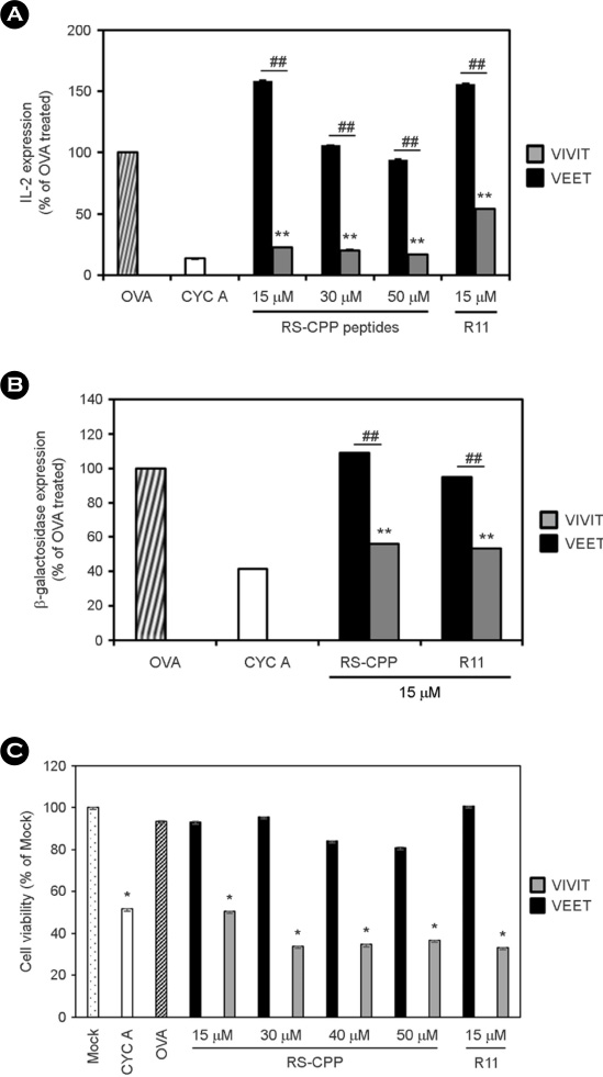
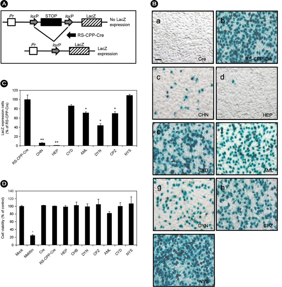
- Cell permeable peptide (CPP) is able to transport itself or conjugated molecules such as nucleotides, peptides, and proteins into cells. Since short peptide of human immunodeficiency virus-1 Tat has been discovered as CPP, it has been continuously studied for their ability to transport heterologous cargoes into cells. In this study, we have focused on the fusion protein of respiratory syncytial virus (RSV), which has six basic amino acids in multi basic furin-dependent cleavage site (MBFCS) required to be cationic CPP. To develop more efficient CPP, the sequence, which linked two MBFCS, was synthesized (called RS-CPP). To assess cell permeable efficiency of RS-CPP or MBFCS, the peptides was conjugated with fluorescein isothiocyanate, and cell permeable efficiency was measured by fluorescence-activated cell sorting. Cell permeability of RS-CPP or MBFCS was increased in a dose-dependent manner, but RS-CPP showed more efficient cell permeability than MBFCS in MDCK, HeLa, Vero E6, and A549 cells. To evaluate whether RS-CPP can transport its conjugated functional peptide (VIVIT) in CD8+ T cell, it was confirmed that IL-2 and β-galactosidase expression were significantly inhibited through selective block of nuclear factor activated T-cell. To investigate endocytic pathways, Cre-mediated DNA recombination (loxP-STOP-loxP-LacZ reporter system) was investigated with divergent endocytosis inhibitors in TE671 cells, and RS-CPP endocytosis is occurred via binding cell surface glycosaminoglycan and clathrin-mediated endocytosis, or macropinocytosis. These results indicated that RS-CPP could be a novel cationic CPP, and it would help understanding for delivery of biologically functional molecules based on viral basic amino acids.
Keyword
MeSH Terms
Figure
Reference
-
1. Heitz F, Morris MC, Divita G. Twenty years of cell-penetrating peptides: from molecular mechanisms to therapeutics. Br J Pharmacol. 2009; 157:195–206.
Article2. Milletti F. Cell-penetrating peptides: classes, origin, and current landscape. Drug Discov Today. 2012; 17:850–860.
Article3. Mäe M, Langel Ü. Cell-penetrating peptides as vectors for peptide, protein and oligonucleotide delivery. Curr Opin Pharmacol. 2006; 6:509–514.
Article4. Rizzuti M, Nizzardo M, Zanetta C, Ramirez A, Corti S. Therapeutic applications of the cell-penetrating HIV-1 Tat peptide. Drug Discov Today. 2015; 20:76–85.
Article5. Wadia JS, Stan RV, Dowdy SF. Transducible TAT-HA fusogenic peptide enhances escape of TAT-fusion proteins after lipid raft macropinocytosis. Nat Med. 2004; 10:310–315.
Article6. Vivès E, Brodin P, Lebleu B. A truncated HIV-1 Tat protein basic domain rapidly translocates through the plasma membrane and accumulates in the cell nucleus. J Biol Chem. 1997; 272:16010–16017.
Article7. Joliot A, Prochiantz A. Transduction peptides: from technology to physiology. Nat Cell Biol. 2004; 6:189–196.
Article8. Futaki S, Suzuki T, Ohashi W, Yagami T, Tanaka S, Ueda K, et al. Arginine-rich peptides An abundant source of membrane-permeable peptides having potential as carriers for intracellular protein delivery. J Biol Chem. 2001; 276:5836–5840.9. Hsieh JT, Zhou J, Gore C, Zimmern P. R11, a novel cell-permeable peptide, as an intravesical delivery vehicle. BJU Int. 2011; 108:1666–1671.
Article10. Tünnemann G, Ter-Avetisyan G, Martin RM, Stöckl M, Herrmann A, Cardoso MC. Live-cell analysis of cell penetration ability and toxicity of oligo-arginines. J Pept Sci. 2008; 14:469–476.
Article11. McLellan JS, Ray WC, Peeples ME. Structure and function of respiratory syncytial virus surface glycoproteins. Curr Top Microbiol Immunol. 2013; 372:83–104.
Article12. González-Reyes L, Ruiz-Argüello MB, García-Barreno B, Calder L, López JA, Albar JP, et al. Cleavage of the human respiratory syncytial virus fusion protein at two distinct sites is required for activation of membrane fusion. Proc Natl Acad Sci U S A. 2001; 98:9859–9864.
Article13. Klimstra WB, Heidner HW, Johnston RE. The furin protease cleavage recognition sequence of Sindbis virus PE2 can mediate virion attachment to cell surface heparan sulfate. J Virol. 1999; 73:6299–6306.
Article14. Rawling J, Cano O, Garcin D, Kolakofsky D, Melero JA. Recombinant Sendai viruses expressing fusion proteins with two furin cleavage sites mimic the syncytial and receptor-independent infection properties of respiratory syncytial virus. J Virol. 2011; 85:2771–2780.
Article15. Noguchi H, Matsushita M, Okitsu T, Moriwaki A, Tomizawa K, Kang S, et al. A new cell-permeable peptide allows successful allogeneic islet transplantation in mice. Nat Med. 2004; 10:305–309.
Article16. Karttunen J, Sanderson S, Shastri N. Detection of rare antigen-presenting cells by the lacZ T-cell activation assay suggests an expression cloning strategy for T-cell antigens. Proc Natl Acad Sci U S A. 1992; 89:6020–6024.
Article17. Fiering S, Northrop JP, Nolan GP, Mattila PS, Crabtree GR, Herzenberg LA. Single cell assay of a transcription factor reveals a threshold in transcription activated by signals emanating from the T-cell antigen receptor. Genes Dev. 1990; 4:1823–1834.
Article18. Chow CW, Rincón M, Davis RJ. Requirement for transcription factor NFAT in interleukin-2 expression. Mol Cell Biol. 1999; 19:2300–2307.
Article19. Xu Y, Liu S, Yu G, Chen J, Chen J, Xu X, et al. Excision of selectable genes from transgenic goat cells by a protein transducible TAT-Cre recombinase. Gene. 2008; 419:70–74.
Article20. Peitz M, Pfannkuche K, Rajewsky K, Edenhofer F. Ability of the hydrophobic FGF and basic TAT peptides to promote cellular uptake of recombinant Cre recombinase: a tool for efficient genetic engineering of mammalian genomes. Proc Natl Acad Sci U S A. 2002; 99:4489–4494.
Article21. Montrose K, Yang Y, Sun X, Wiles S, Krissansen GW. Xentry, a new class of cell-penetrating peptide uniquely equipped for delivery of drugs. Sci Rep. 2013; 3:1661.
Article22. Fang SL, Fan TC, Fu HW, Chen CJ, Hwang CS, Hung TJ, et al. A novel cell-penetrating peptide derived from human eosinophil cationic protein. PLoS One. 2013; 8:e57318.
Article23. Hakansson S, Jacobs A, Caffrey M. Heparin binding by the HIV-1 tat protein transduction domain. Protein Sci. 2001; 10:2138–2139.
Article24. Richard JP, Melikov K, Brooks H, Prevot P, Lebleu B, Chernomordik LV. Cellular uptake of unconjugated TAT peptide involves clathrin-dependent endocytosis and heparan sulfate receptors. J Biol Chem. 2005; 280:15300–15306.
Article
- Full Text Links
- Actions
-
Cited
- CITED
-
- Close
- Share
- Similar articles
-
- Cloning of Novel Epidermal Growth Factor (EGF) Plasmid for Gene Therapy on Diabetic Foot Ulcer
- A basic study for respiratory sybcytial virus detection using polymerase chain reaction
- Respiratory Syncytial Virus Infections and Application of Nested Reverse Transcription-Polymerase Chain Reaction
- Recovery of respiratory syncytial virus, adenovirus, influenza virus , and parainfluenza virus from nasopharyngeal aspirates from children with acute respiratory tract infections
- Immunogenicity and Protective Efficacy of a Dual Subunit Vaccine Against Respiratory Syncytial Virus and Influenza Virus


