J Vet Sci.
2017 Aug;18(S1):361-370. 10.4142/jvs.2017.18.S1.361.
Generation, characterization, and application in serodiagnosis of recombinant swine vesicular disease virus-like particles
- Affiliations
-
- 1National Centre for Foreign Animal Disease, Winnipeg, MB R3E 3M4, Canada. Ming.yang@inspection.gc.ca
- KMID: 2412561
- DOI: http://doi.org/10.4142/jvs.2017.18.S1.361
Abstract
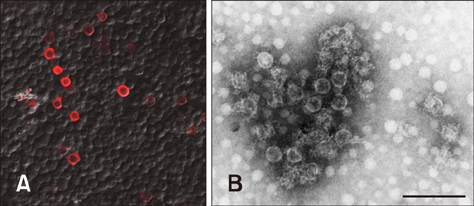
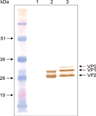
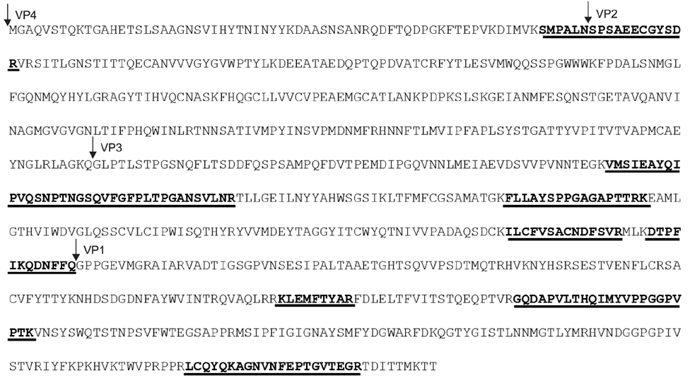
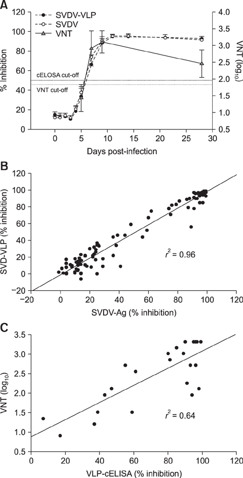
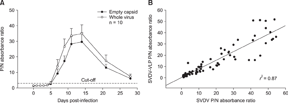
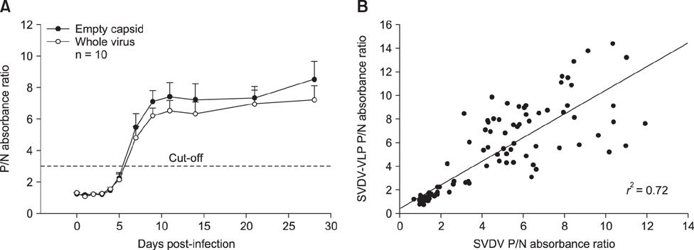
- Swine vesicular disease (SVD) is a highly contagious viral disease that causes vesicular disease in pigs. The importance of the disease is due to its indistinguishable clinical signs from those of foot-and-mouth disease, which prevents international trade of swine and related products. SVD-specific antibody detection via an enzyme-linked immunosorbent assay (ELISA) is the most versatile and commonly used method for SVD surveillance and export certification. Inactivated SVD virus is the commonly used antigen in SVD-related ELISA. A recombinant SVD virus-like particle (VLP) was generated by using a Bac-to-Bac baculovirus expression system. Results of SVD-VLP analyses from electron microscopy, western blotting, immunofluorescent assay, and mass spectrometry showed that the recombinant SVD-VLP morphologically resemble authentic SVD viruses. The SVD-VLP was evaluated as a replacement for inactivated whole SVD virus in competitive and isotype-specific ELISAs for the detection of antibodies against SVD virus. The recombinant SVD-VLP assay produced results similar to those from inactivated whole virus antigen ELISA. The VLP-based ELISA results were comparable to those from the virus neutralization test for antibody detection in pigs experimentally inoculated with SVD virus. Use of the recombinant SVD-VLP is a safe and valuable alternative to using SVD virus antigen in diagnostic assays.
Keyword
MeSH Terms
Figure
Reference
-
1. Basavappa R, Syed R, Flore O, Icenogle JP, Filman DJ, Hogle JM. Role and mechanism of the maturation cleavage of VP0 in poliovirus assembly: structure of the empty capsid assembly intermediate at 2.9 Å resolution. Protein Sci. 1994; 3:1651–1669.
Article2. Brocchi E, Berlinzani A, Gamba D, De Simone F. Development of two novel monoclonal antibody-based ELISAs for the detection of antibodies and the identification of swine isotypes against swine vesicular disease virus. J Virol Methods. 1995; 52:155–167.
Article3. De Clercq K. Reduction of singleton reactors against swine vesicular disease virus by a combination of virus neutralisation test, monoclonal antibody-based competitive ELISA and isotype specific ELISA. J Virol Methods. 1998; 70:7–18.
Article4. Dekker A, van Hemert-Kluitenberg F, Baars C, Terpstra C. Isotype specific ELISAs to detect antibodies against swine vesicular disease virus and their use in epidemiology. Epidemiol Infect. 2002; 128:277–284.
Article5. Hammond GW, Hazelton PR, Chuang I, Klisko B. Improved detection of viruses by electron microscopy after direct ultracentrifuge preparation of specimens. J Clin Microbiol. 1981; 14:210–221.
Article6. Heckert RA, Brocchi E, Berlinzani A, Mackay DK. An international comparative analysis of a competitive ELISA for the detection of antibodies to swine vesicular disease virus. J Vet Diagn Invest. 1998; 10:295–297.
Article7. Inoue T, Suzuki T, Sekiguchi K. The complete nucleotide sequence of swine vesicular disease virus. J Gen Virol. 1989; 70:919–934.
Article8. Inoue T, Yamaguchi S, Kanno T, Sugita S, Saeki T. The complete nucleotide sequence of a pathogenic swine vesicular disease virus isolated in Japan (J1'73) and phylogenetic analysis. Nucleic Acids Res. 1993; 21:3896.
Article9. Jore J, De Geus B, Jackson RJ, Pouwels PH, Enger-Valk BE. Poliovirus protein 3CD is the active protease for processing of the precursor protein P1 in vitro. J Gen Virol. 1988; 69:1627–1636.
Article10. Karakas M, Palfi V. [Diagnostic difficulties during the serological examination of swine vesicular disease]. Magy Allatorvosok Lapja. 1996; 51:595–597. Hungarian.11. Ko YJ, Choi KS, Nah JJ, Paton DJ, Oem JK, Wilsden G, Kang SY, Jo NI, Lee JH, Kim JH, Lee HW, Park JM. Noninfectious virus-like particle antigen for detection of swine vesicular disease virus antibodies in pigs by enzyme-linked immunosorbent assay. Clin Diagn Lab Immunol. 2005; 12:922–929.
Article12. Lin F, Kitching RP. Swine vesicular disease: an overview. Vet J. 2000; 160:192–201.
Article13. Nijhar SK, Mackay DK, Brocchi E, Ferris NP, Kitching RP, Knowles NJ. Identification of neutralizing epitopes on a European strain of swine vesicular disease virus. J Gen Virol. 1999; 80:277–282.
Article14. Racaniello VR. Picornaviridae: the viruses and their replication. In : Knipe DM, Howley PM, editors. Fields Virology. 5th ed. Philadelphia: Lippincott Williams & Wilkins;2007. p. 795–838.15. Seechurn P, Knowles NJ, McCauley JW. The complete nucleotide sequence of a pathogenic swine vesicular disease virus. Virus Res. 1990; 16:255–274.
Article16. Vratskikh O, Stiasny K, Zlatkovic J, Tsouchnikas G, Jarmer J, Karrer U, Roggendorf M, Roggendorf H, Allwinn R, Heinz FX. Dissection of antibody specificities induced by yellow fever vaccination. PLoS Pathog. 2013; 9:e1003458.
Article17. World Organisation for Animal Health (OIE). Swine vesicular disease. OIE Biological Standards Commission. Manual of Diagnostic Tests and Vaccines for Terrestrial Animals. Volume 2:6th ed. Paris: Office International des Epizooties;2008. p. 1139–1145.18. Yang M, Clavijo A. Monoclonal antibody against swine vesicular disease virus. Hybridoma. 2008; 27:325.
Article19. Yang M, Clavijo A, Suarez-Banmann R, Avalo R. Production and characterization of two serotype independent monoclonal antibodies against foot-and-mouth disease virus. Vet Immunol Immunopathol. 2007; 115:126–134.
Article20. Ypma-Wong MF, Dewalt PG, Johnson VH, Lamb JG, Semler BL. Protein 3CD is the major poliovirus proteinase responsible for cleavage of the p1 capsid precursor. Virology. 1988; 166:265–270.
Article
- Full Text Links
- Actions
-
Cited
- CITED
-
- Close
- Share
- Similar articles
-
- Progress and hurdles in the development of influenza virus-like particle vaccines for veterinary use
- Characterization of the Recombinant Proteins of Porcine Circovirus Type2 Field Isolate Expressed in the Baculovirus System
- Development of a novel enzyme-linked immunosorbent assay to detect anti-IgG against swine hepatitis E virus
- Establishment and characterization of an infectious cDNA clone of a classical swine fever virus LOM strain
- Molecular Approach to Allergy Diagnosis and Therapy

