Clin Orthop Surg.
2016 Dec;8(4):373-378. 10.4055/cios.2016.8.4.373.
Biomechanical Analysis of a Novel Wedge Locking Plate in a Porcine Tibial Model
- Affiliations
-
- 1Department of Orthopedic Surgery, Seoul Paik Hospital, University of Inje College of Medicine, Seoul, Korea.
- 2Department of Biomedical Engineering, Inje University, Gimhae, Korea.
- 3Trauma & Orthopaedics, Medway NHS Trust, Kent, UK.
- 4Department of Orthopaedic Surgery, Konkuk University School of Medicine, Seoul, Korea. boram107@hanmail.net
- KMID: 2412318
- DOI: http://doi.org/10.4055/cios.2016.8.4.373
Abstract
- BACKGROUND
The purpose of this study was to analyze biomechanical properties of a novel wedge locking plate in medial open wedge high tibial osteotomy (OWHTO) in a porcine tibial model.
METHODS
A uniform 8-mm OWHTO was performed in 12 porcine tibiae. Six of them were subsequently fixed with the plate without a wedge, whereas the other 6 were additionally reinforced with a metal wedge of 8 mm. Biomechanical properties (stiffness, displacement of the osteotomy gap, and failure load) were evaluated under axial load. The different modes of failure were also investigated.
RESULTS
The plate showed an axial stiffness of 2,457 ± 450 N/mm with a wedge and 1,969 ± 874 N/mm without a wedge. The maximum failure load was 5,380 ± 952 N with a wedge and 4,354 ± 607 N without a wedge. The plate with a wedge had a significantly greater failure load and significantly less displacement of medial gap at failure than that without a wedge (p = 0.041 and p = 0.002, respectively). The axial stiffness was not different between the two types of fixation. Most failures were caused by lateral cortex breakage and there was no implant failure.
CONCLUSIONS
The novel wedge locking plate showed excellent biomechanical properties and an additional wedge provided significant improvement. This plate can be a good fixation method for OWHTO.
Keyword
MeSH Terms
Figure
-

Fig. 1 A novel wedge locking plate.
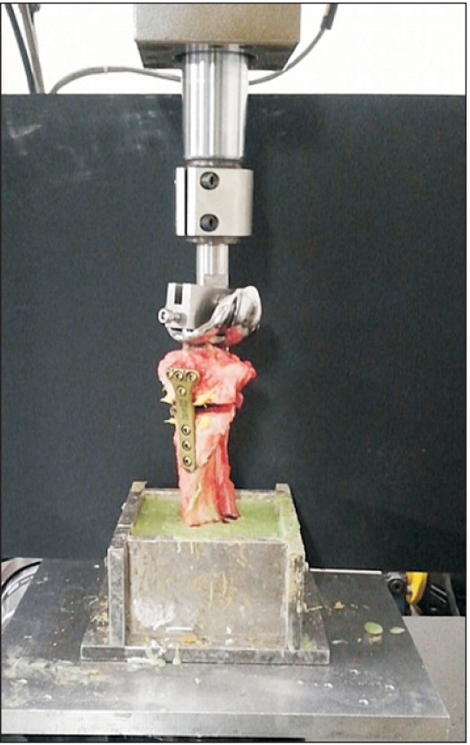
Fig. 2 Testing model mounted on MTS (MTS 858 Bionix; MTS System Co.). A prosthesis for total knee replacement was used for the axial load. The specimen was embedded in polyurethane casting resin and mounted in a fixture.
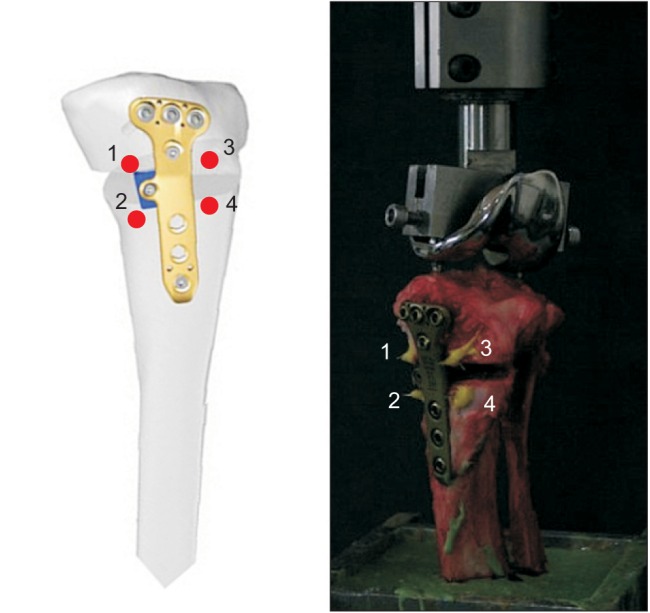
Fig. 3 Schematic drawing of four markers for the displacement measurement (ventral and dorsal sides of both proximal and distal tibial surfaces). 1 and 2 are for posteromedial gap and 3 and 4 are for anterolateral gap.
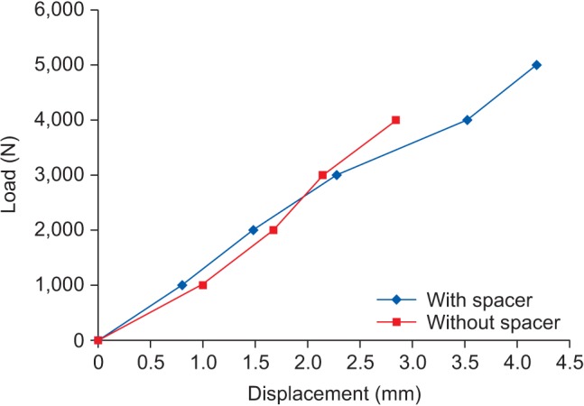
Fig. 4 A graph for the medial gap displacement. At failure load, significant difference was shown between two groups.
Reference
-
1. Saito T, Kumagai K, Akamatsu Y, Kobayashi H, Kusayama Y. Five- to ten-year outcome following medial opening-wedge high tibial osteotomy with rigid plate fixation in combination with an artificial bone substitute. Bone Joint J. 2014; 96(3):339–344. PMID: 24589788.
Article2. Floerkemeier S, Staubli AE, Schroeter S, Goldhahn S, Lobenhoffer P. Outcome after high tibial open-wedge osteotomy: a retrospective evaluation of 533 patients. Knee Surg Sports Traumatol Arthrosc. 2013; 21(1):170–180. PMID: 22744433.
Article3. Spahn G, Muckley T, Kahl E, Hofmann GO. Biomechanical investigation of different internal fixations in medial opening-wedge high tibial osteotomy. Clin Biomech (Bristol, Avon). 2006; 21(3):272–278.
Article4. Agneskirchner JD, Freiling D, Hurschler C, Lobenhoffer P. Primary stability of four different implants for opening wedge high tibial osteotomy. Knee Surg Sports Traumatol Arthrosc. 2006; 14(3):291–300. PMID: 16284740.
Article5. Schroter S, Gonser CE, Konstantinidis L, Helwig P, Albrecht D. High complication rate after biplanar open wedge high tibial osteotomy stabilized with a new spacer plate (Position HTO plate) without bone substitute. Arthroscopy. 2011; 27(5):644–652. PMID: 21663721.6. Nelissen EM, van Langelaan EJ, Nelissen RG. Stability of medial opening wedge high tibial osteotomy: a failure analysis. Int Orthop. 2010; 34(2):217–223. PMID: 19189104.
Article7. Jung WH, Chun CW, Lee JH, Ha JH, Kim JH, Jeong JH. Comparative study of medial opening-wedge high tibial osteotomy using 2 different implants. Arthroscopy. 2013; 29(6):1063–1071. PMID: 23623294.
Article8. Valkering KP, van den Bekerom MP, Kappelhoff FM, Albers GH. Complications after tomofix medial opening wedge high tibial osteotomy. J Knee Surg. 2009; 22(3):218–225. PMID: 19634725.
Article9. Han SB, Bae JH, Lee SJ, et al. Biomechanical properties of a new anatomical locking metal block plate for opening wedge high tibial osteotomy: uniplane osteotomy. Knee Surg Relat Res. 2014; 26(3):155–161. PMID: 25229045.
Article10. Andriacchi TP, Andersson GB, Fermier RW, Stern D, Galante JO. A study of lower-limb mechanics during stair-climbing. J Bone Joint Surg Am. 1980; 62(5):749–757. PMID: 7391098.
Article11. Kuster MS, Wood GA, Stachowiak GW, Gachter A. Joint load considerations in total knee replacement. J Bone Joint Surg Br. 1997; 79(1):109–113. PMID: 9020457.
Article12. Morrison JB. The mechanics of the knee joint in relation to normal walking. J Biomech. 1970; 3(1):51–61. PMID: 5521530.
Article13. Stoffel K, Stachowiak G, Kuster M. Open wedge high tibial osteotomy: biomechanical investigation of the modified Arthrex Osteotomy Plate (Puddu Plate) and the TomoFix Plate. Clin Biomech (Bristol, Avon). 2004; 19(9):944–950.
Article14. Tetsumura S, Fujita A, Nakajima M, Abe M. Biomechanical comparison of different fixation methods on the tibial side in anterior cruciate ligament reconstruction: a biomechanical study in porcine tibial bone. J Orthop Sci. 2006; 11(3):278–282. PMID: 16721530.
Article15. Yoo JH, Seong SC, Lee S, et al. Rigid stepped plate for internal fixation for high tibial osteotomy. Orthopedics. 2009; 32(10):DOI: 10.3928/01477447-20090818-08.
Article
- Full Text Links
-
- Actions
-
Cited
- CITED
-
- Close
- Share
-
- Similar articles
-
- A Short Term Follow-up of Open Wedge High Tibial Osteotomyusing Locking Compression Plate(R)
- Minimally Invasive Percutaneous Plate Stabilization Using a Medial Locking Plate for Proximal Tibial Fractures: Technical Note
- Back out of Locking Pin with Hinge Fracture after High Tibial Osteotomy
- Biomechanical Properties of a New Anatomical Locking Metal Block Plate for Opening Wedge High Tibial Osteotomy: Uniplane Osteotomy
- Biomechanical Analysis of Operative Methods in the Treatment of Extra-Articular Fracture of the Proximal Tibia
