Effects of Different Angles of the Traction Table on Lumbar Spine Ligaments: A Finite Element Study
- Affiliations
-
- 1Department of Biomedical Engineering, Faculty of Engineering, University of Isfahan, Isfahan, Iran. n.jamshidi@eng.ui.ac.ir
- KMID: 2412249
- DOI: http://doi.org/10.4055/cios.2017.9.4.480
Abstract
- BACKGROUND
The traction bed is a noninvasive device for treating lower back pain caused by herniated intervertebral discs. In this study, we investigated the impact of the traction bed on the lower back as a means of increasing the disc height and creating a gap between facet joints.
METHODS
Computed tomography (CT) images were obtained from a female volunteer and a three-dimensional (3D) model was created using software package MIMICs 17.0. Afterwards, the 3D model was analyzed in an analytical software (Abaqus 6.14). The study was conducted under the following traction loads: 25%, 45%, 55%, and 85% of the whole body weight in different angles.
RESULTS
Results indicated that the loading angle in the L3-4 area had 36.8%, 57.4%, 55.32%, 49.8%, and 52.15% effect on the anterior longitudinal ligament, posterior longitudinal ligament, intertransverse ligament, interspinous ligament, and supraspinous ligament, respectively. The respective values for the L4-5 area were 32.3%, 10.6%, 53.4%, 56.58%, and 57.35%. Also, the body weight had 63.2%, 42.6%, 44.68%, 50.2%, and 47.85% effect on the anterior longitudinal ligament, posterior longitudinal ligament, intertransverse ligament, interspinous ligament, and supraspinous ligament, respectively. The respective values for the L4-5 area were 67.7%, 89.4%, 46.6%, 43.42% and 42.65%. The authenticity of results was checked by comparing with the experimental data.
CONCLUSIONS
The results show that traction beds are highly effective for disc movement and lower back pain relief. Also, an optimal angle for traction can be obtained in a 3D model analysis using CT or magnetic resonance imaging images. The optimal angle would be different for different patients and thus should be determined based on the decreased height of the intervertebral disc, weight and height of patients.
Keyword
MeSH Terms
-
Adult
Biomechanical Phenomena
Body Weight
Computer Simulation
Elasticity
Female
Finite Element Analysis
Humans
Imaging, Three-Dimensional
Intervertebral Disc/*diagnostic imaging/physiology
Longitudinal Ligaments/*diagnostic imaging/physiology
Lumbar Vertebrae/*diagnostic imaging
Patient Positioning
Tomography, X-Ray Computed
*Traction/instrumentation
Viscosity
Figure
-
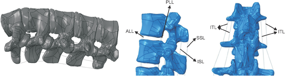
Fig. 1 Lumbar spine model with ligaments. ALL: anterior longitudinal ligament, PLL: posterior longitudinal ligament, SSL: supraspinous ligament, ISL: interspinous ligament, ITL: intertransverse ligament.

Fig. 2 (A) Disc model with two fibrosus parts of nucleus pulposus (NP) and annulus fibrosis (AF). (B) Maxwell model for viscoelastic materials.11)
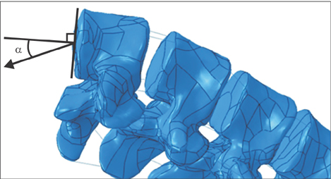
Fig. 3 Load and angle apply method in Taguchi design of experiment.
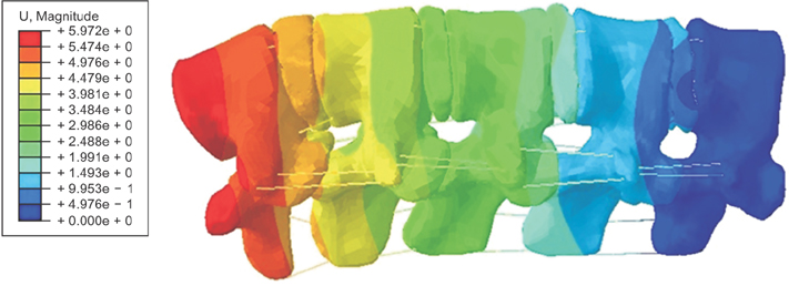
Fig. 4 Relative displacements of intervertebral discs (mm) under 45% of body weight loading.
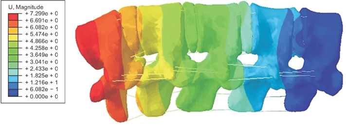
Fig. 5 Relative displacements of intervertebral discs (mm) under 55% of body weight loading.
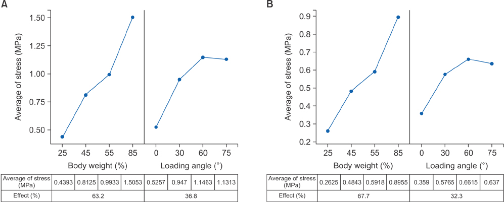
Fig. 6 The results of Taguchi design of experiment on anterior longitudinal ligament in the L3–4 area (A) and L4–5 area (B).

Fig. 7 The results of Taguchi design of experiment on posterior longitudinal ligament in the L3–4 area (A) and L4–5 area (B).
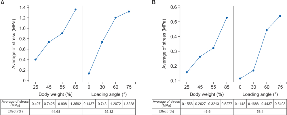
Fig. 8 The results of Taguchi design of experiment on intertransverse ligament in the L3–4 area (A) and L4–5 area (B).

Fig. 9 The results of Taguchi design of experiment on interspinous ligament in the L3–4 area (A) and L4–5 area (B).
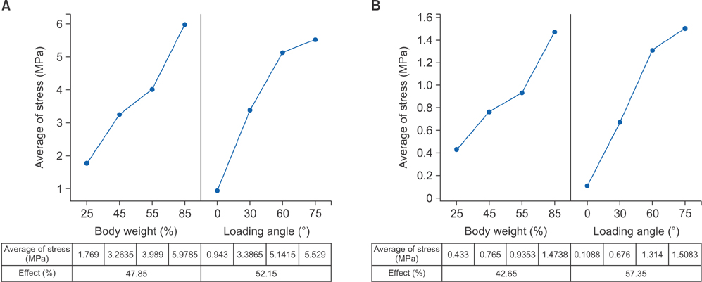
Fig. 10 The results of Taguchi design of experiment on supraspinous ligament in the L3–4 area (A) and L4–5 area (B).

Fig. 11 Comparison between results of this study and a clinical study for 45% of body weight loading.
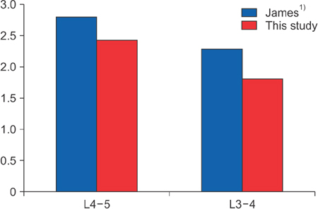
Fig. 12 Comparison between results of this study and a clinical study for 55% of body weight loading.
Reference
-
1. James M. Mechanically assisted spinal mobilization. In : Stude DE, editor. Spinal rehabilitation. New York, NY: McGraw-Hill Professional;1999. p. 417–442.2. Kamanli A, Karaca-Acet G, Kaya A, Koc M, Yildirim H. Conventional physical therapy with lumbar traction: clinical evaluation and magnetic resonance imaging for lumbar disc herniation. Bratisl Lek Listy. 2010; 111(10):541–544.3. Skrzypiec DM, Pollintine P, Przybyla A, Dolan P, Adams MA. The internal mechanical properties of cervical intervertebral discs as revealed by stress profilometry. Eur Spine J. 2007; 16(10):1701–1709.
Article4. Pitzen T, Geisler F, Matthis D, et al. A finite element model for predicting the biomechanical behaviour of the human lumbar spine. Control Eng Pract. 2002; 10(1):83–90.
Article5. Ibarz E, Herrera A, Mas Y, et al. Development and kinematic verification of a finite element model for the lumbar spine: application to disc degeneration. Biomed Res Int. 2013; 2013:705185. DOI: 10.1155/2013/705185.
Article6. Clarke J, van Tulder M, Blomberg S, de Vet H, van der Heijden G, Bronfort G. Traction for low back pain with or without sciatica: an updated systematic review within the framework of the Cochrane collaboration. Spine (Phila Pa 1976). 2006; 31(14):1591–1599.
Article7. Meijer GJ, Homminga J, Hekman EE, Veldhuizen AG, Verkerke GJ. The effect of three-dimensional geometrical changes during adolescent growth on the biomechanics of a spinal motion segment. J Biomech. 2010; 43(8):1590–1597.
Article8. Park WM, Kim K, Kim YH. Biomechanical analysis of two-step traction therapy in the lumbar spine. Man Ther. 2014; 19(6):527–533.
Article9. Wang JL, Parnianpour M, Shirazi-Adl A, Engin AE. Viscoelastic finite-element analysis of a lumbar motion segment in combined compression and sagittal flexion: effect of loading rate. Spine (Phila Pa 1976). 2000; 25(3):310–318.
Article10. Shin G, Mirka GA, Loboa EG. Viscoelastic responses of the lumbar spine during prolonged stooping. Proc Hum Factors Ergon Soc Annu Meet. 2005; 49(14):1269–1273.
Article11. ANSYS. ANSYS academic research, released 15, help system: coupled-field analysis guide. Canonsburg, PA: ANSYS Inc.;2015.12. Dassault Systemes. ABAQUS 6.14 analysis user's manual: online documentation help. Velizy-Villacoublay, France: Dassault Systemes;2014.13. del Palomar AP, Calvo B, Doblare M. An accurate finite element model of the cervical spine under quasi-static loading. J Biomech. 2008; 41(3):523–531.
Article14. Beattie PF, Nelson RM, Michener LA, Cammarata J, Donley J. Outcomes after a prone lumbar traction protocol for patients with activity-limiting low back pain: a prospective case series study. Arch Phys Med Rehabil. 2008; 89(2):269–274.
Article15. Horseman I, Morningstar MW. Radiographic disk height increase after a trial of multimodal spine rehabilitation and vibration traction: a retrospective case series. J Chiropr Med. 2008; 7(4):140–145.
Article16. Kurutzne KM, Bene E, Lovas A, Molnar P, Monori E. Biomechanical experiments for measuring traction lengthening of the lumbar spine during weight bath therapy. Orv Hetil. 2002; 143(13):673–684.
- Full Text Links
-
- Actions
-
Cited
- CITED
-
- Close
- Share
-
- Similar articles
-
- Comparative Finite Element Analysis of Lumbar Cortical Screws and Pedicle Screws in Transforaminal and Posterior Lumbar Interbody Fusion
- Analysis of Biomechanical Properties of Whole Cervical Spine under Static Loading with 3-D Finite Element Model
- Analysis of Compression Behavior on Intervertebral Disc L4-5 in Pedicle Screw System Instrumented Lumbar Spine under Follower Load
- Stress Analysis of the Lumbar Spine under Dynamic Loading Condition with 3 - D Nonlinear Finite Element Model
- Analysis of Compression Behavior in Lumbar Spine Under Simple Vertical Load vs Follower Load
