Thickening Ligamentum Flavum Mimicking Tumor in the Epidural Space of the Cervical Spine
- Affiliations
-
- 1Department of Neurosurgery, Pusan National University Yangsan Hospital, Pusan National University School of Medicine, Yangsan, Korea. md6576@naver.com
- KMID: 2410922
- DOI: http://doi.org/10.13004/kjnt.2018.14.1.43
Abstract
- In patients with tumors and spinal cord lesions, inflammation and tissue infection can result in mass effect detection on imaging. As a result, surgical biopsy procedures are often performed on the lesions. We report a rare case in which the thickening ligamentum flavum (LF) appeared to be a tumor in the epidural space of the cervical spine based on imaging findings. A 52-year-old man visited our outpatient clinic with severe shoulder pain and radicular pain in his right arm that had developed gradually after a traffic accident two months earlier. Magnetic resonance imaging of the cervical spine revealed an extradural mass at the cervicothoracic junction level. Suspecting a tumor, spinal decompression surgery was performed and a biopsy of the mass was obtained. At the time of surgery, the LF was thick and compressed the spinal cord. After successful removal of the LF, the spinal cord appeared normal. Histopathological examination confirmed the mass as the LF. The patient was discharged without pain or weakness two weeks postoperatively. This case demonstrated that when the LF of the cervicothoracic junction is thickened, it may be misdiagnosed as a cervical spine tumor compressing the spinal cord.
MeSH Terms
Figure
-
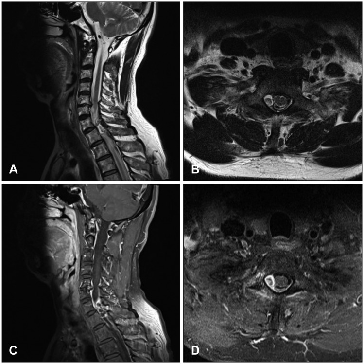
FIGURE 1 Preoperative T2-weighted magnetic resonance images (MRIs): (A) sagittal and (B) axial views. T1-weighted MRIs with gadolinium: (C) sagittal and (D) axial views. The images revealed an extradural mass at the right epidural space of the C7-T1 level.

FIGURE 2 (A) Intraoperative photograph showing the hard and stiff epidural mass (arrow) compressed the spinal cord. (B) After careful removal of the epidural mass using a dissector and punch, the spinal cord appeared normal.
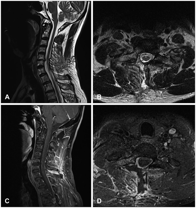
FIGURE 3 Postoperative T2-weighted magnetic resonance images (MRIs): (A) sagittal and (B) axial views. T1-weighted MRIs with gadolinium: (C) sagittal and (D) axial views. The images revealed remarkable resolution of the mass after right hemilaminectomy.
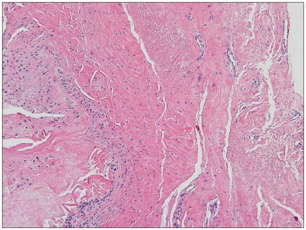
FIGURE 4 Microscopic photograph of the section from the resected mass compressing the spinal cord, original magnification ×100. The resected mass was a ligamentum flavum, containing elastic fibers with irregular arrangement, reduced elastic components, and increased collagen tissue.
Reference
-
1. Grenier N, Kressel HY, Schiebler ML, Grossman RI, Dalinka MK. Normal and degenerative posterior spinal structures: MR imaging. Radiology. 1987; 165:517–525. PMID: 3659376.
Article2. Ivancic PC, Coe MP, Ndu AB, Tominaga Y, Carlson EJ, Rubin W, et al. Dynamic mechanical properties of intact human cervical spine ligaments. Spine J. 2007; 7:659–665. PMID: 17998125.
Article3. Runge VM, Lee C, Iten AL, Williams NM. Contrast-enhanced magnetic resonance imaging in a spinal epidural tumor model. Invest Radiol. 1997; 32:589–595. PMID: 9342117.
Article4. Sairyo K, Biyani A, Goel V, Leaman D, Booth R Jr, Thomas J, et al. Pathomechanism of ligamentum flavum hypertrophy: a multidisciplinary investigation based on clinical, biomechanical, histologic, and biologic assessments. Spine (Phila Pa 1976). 2005; 30:2649–2656. PMID: 16319751.
Article5. Sakamaki T, Sairyo K, Sakai T, Tamura T, Okada Y, Mikami H. Measurements of ligamentum flavum thickening at lumbar spine using MRI. Arch Orthop Trauma Surg. 2009; 129:1415–1419. PMID: 19280205.
Article6. Sayit E, Daubs MD, Aghdasi B, Montgomery SR, Inoue H, Wang CJ, et al. Dynamic changes of the ligamentum flavum in the cervical spine assessed with kinetic magnetic resonance imaging. Global Spine J. 2013; 3:69–74. PMID: 24436854.
Article7. Taylor AR. The mechanism of injury to the spinal cord in the neck without damage to vertebral column. J Bone Joint Surg Br. 1951; 33B:543–547.
- Full Text Links
-
- Actions
-
Cited
- CITED
-
- Close
- Share
-
- Similar articles
-
- Anatomic Variations of Cervical and High Thoracic Ligamentum Flavum
- Comparison of Methods to Confirm the Cervical Epidural Space
- Drip infusion method as a useful indicator for identification of the epidural space
- Ossified Ligamentum Flavum causing Cervical Myelopathy
- Cervical Radiculomyelopathy due to Calcification of the Ligamentum Flavum
