The Potential Role of Grid-Like Software in Bedside Chest Radiography in Improving Image Quality and Dose Reduction: An Observer Preference Study
- Affiliations
-
- 1Department of Radiology, Seoul National University College of Medicine, and Institute of Radiation Medicine, Seoul National University Medical Research Center, Seoul 03080, Korea. jmgoo@plaza.snu.ac.kr
- 2Department of Radiology, Konkuk University Medical Center, Konkuk University School of Medicine, Seoul 05030, Korea.
- 3Department of Radiology, Institute of Medical Science, Research Institute of Clinical Medicine, Chonbuk National University Medical School and Hospital, Jeonju 54907, Korea.
- 4Cancer Research Institute, Seoul National University College of Medicine, Seoul 03080, Korea.
- KMID: 2410824
- DOI: http://doi.org/10.3348/kjr.2018.19.3.526
Abstract
OBJECTIVE
To compare the observer preference of image quality and radiation dose between non-grid, grid-like, and grid images.
MATERIALS AND METHODS
Each of the 38 patients underwent bedside chest radiography with and without a grid. A grid-like image was generated from a non-grid image using SimGrid software (Samsung Electronics Co. Ltd.) employing deep-learning-based scatter correction technology. Two readers recorded the preference for 10 anatomic landmarks and the overall appearance on a five-point scale for a pair of non-grid and grid-like images, and a pair of grid-like and grid images, respectively, which were randomly presented. The dose area product (DAP) was also recorded. Wilcoxon's rank sum test was used to assess the significance of preference.
RESULTS
Both readers preferred grid-like images to non-grid images significantly (p < 0.001); with a significant difference in terms of the preference for grid images to grid-like images (p = 0.317, 0.034, respectively). In terms of anatomic landmarks, both readers preferred grid-like images to non-grid images (p < 0.05). No significant differences existed between grid-like and grid images except for the preference for grid images in proximal airways by two readers, and in retrocardiac lung and thoracic spine by one reader. The median DAP were 1.48 (range, 1.37-2.17) dGy*cm2 in grid images and 1.22 (range, 1.11-1.78) dGy*cm2 in grid-like images with a significant difference (p < 0.001).
CONCLUSION
The SimGrid software significantly improved the image quality of non-grid images to a level comparable to that of grid images with a relatively lower level of radiation exposure.
Keyword
Figure
-
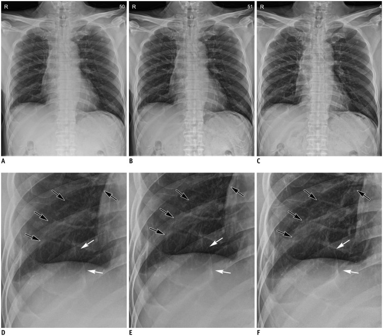
Fig. 1 Comparison of (A, D) non-grid, (B, E) grid-like, and (C, F) grid images.A-C. Chest radiography of 57-year-old male patient who underwent multiple-wedge resection in both lungs for metastasis associated with undifferentiated pleomorphic sarcoma. Note superior image contrast of grid-like image to non-grid image, and similarity in contrast appearance between grid-like and grid images. D-F. Magnified non-grid, grid-like and grid images: surgical materials (black arrows) and multiple linear opacities (white arrows) are clearly demonstrated in grid-like image, as well as grid image.
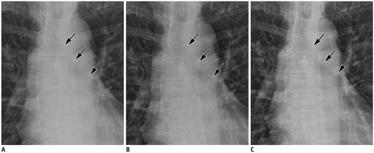
Fig. 2 Magnified anteroposterior chest radiographs of 59-year-old female with ovarian cancer.Compared with (A) non-grid image, delineation of trachea and left main bronchus (arrows) is improved in (B) grid-like image, as well as (C) grid image.
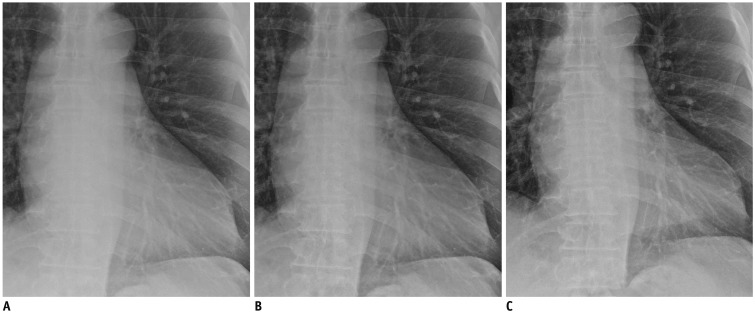
Fig. 3 Magnified anteroposterior chest radiographs of 63-year-old male with hepatocellular carcinoma.Compared with (A) non-grid image, delineation of vascular shadows in retrocardiac area is improved in (B) grid-like image, as well as (C) grid image.
Reference
-
1. Rubinowitz AN, Siegel MD, Tocino I. Thoracic imaging in the ICU. Crit Care Clin. 2007; 23:539–573. PMID: 17900484.
Article2. Eisenhuber E, Schaefer-Prokop CM, Prosch H, Schima W. Bedside chest radiography. Respir Care. 2012; 57:427–443. PMID: 22391269.
Article4. Kawamura T, Naito S, Okano K, Yamada M. Improvement in image quality and workflow of x-ray examinations using a new image processing method, “Virtual Grid Technology”. Web site. 2015. Accessed July 13, 2017. http://www.fujifilm.com/about/research/report/060/pdf/index/ff_rd060_004_en.pdf.5. Mentrup D, Specht S, Spieckermann J, Zorba G. Gridless imaging with SkyFlow: time savings and workflow improvements. 2015. 6. Accessed July 13, 2017. https://www.usa.philips.com/b-dam/b2bhc/us/topics/radiology-patience-experience/SkyFlowwhitepaper-452299111471_LR.PDF.6. Mentrup D, Jockel S, Menser B, Neitzel U. Iterative scatter correction for grid-less bedside chest radiography: performance for a chest phantom. Radiat Prot Dosimetry. 2016; 169:308–312. PMID: 26487750.
Article7. Renger B, Brieskorn C, Toth V, Mentrup D, Jockel S, Lohöfer F, et al. Evaluation of dose reduction potentials of a novel scatter correction software for bedside chest X-ray imaging. Radiat Prot Dosimetry. 2016; 169:60–67. PMID: 26977074.
Article8. Goo JM, Im JG, Kim JH, Seo JB, Kim TS, Shine SJ, et al. Digital chest radiography with a selenium-based flat-panel detector versus a storage phosphor system: comparison of soft-copy images. AJR Am J Roentgenol. 2000; 175:1013–1018. PMID: 11000155.9. Floyd CE Jr, Baker JA, Lo JY, Ravin CE. Measurement of scatter fractions in clinical bedside radiography. Radiology. 1992; 183:857–861. PMID: 1584947.
Article10. Anderson DW. Introduction of grids to mobile ICU radiography in a teaching hospital. Br J Radiol. 2006; 79:315–318. PMID: 16585724.
Article11. Moore CS, Wood TJ, Avery G, Balcam S, Needler L, Smith A, et al. Investigating the use of an antiscatter grid in chest radiography for average adults with a computed radiography imaging system. Br J Radiol. 2015; 88:20140613. PMID: 25571914.
Article12. Scott AW, Gauntt DM, Yester MV, Barnes GT. High-ratio grid considerations in mobile chest radiography. Med Phys. 2012; 39:3142–3153. PMID: 22755699.
Article13. Uffmann M, Schaefer-Prokop C. Digital radiography: the balance between image quality and required radiation dose. Eur J Radiol. 2009; 72:202–208. PMID: 19628349.
Article14. Gauntt DM, Barnes GT. An automatic and accurate x-ray tube focal spot/grid alignment system for mobile radiography: system description and alignment accuracy. Med Phys. 2010; 37:6402–6410. PMID: 21302797.15. MacMahon H, Yasillo NJ, Carlin M. Laser alignment system for high-quality portable radiography. Radiographics. 1992; 12:111–120. PMID: 1734456.
Article16. Chae KJ, Goo JM, Ahn SY, Yoo JY, Yoon SH. Application of deconvolution algorithm of point spread function in improving image quality: an observer preference study on chest radiography. Korean J Radiol. 2018; 19:147–152. PMID: 29354011.
Article17. Lee K, Lee K, Lee S, Kim H. Workflow improvement with Samsung SimGrid SW. Web site. 2016. 2. 22. Accessed July 13, 2017. https://www.samsungmedicalsolution.com/en/common/whitePaper.
- Full Text Links
-
- Actions
-
Cited
- CITED
-
- Close
- Share
-
- Similar articles
-
- Application of Deconvolution Algorithm of Point Spread Function in Improving Image Quality: An Observer Preference Study on Chest Radiography
- Objective and Quantitative Evaluation of Image Quality Using Fuzzy Integral: Phantom Study
- Relationship of Image Quality and Radiation Dose for Chest Radiography in the Medical Institutions for Pneumoconiosis: A Comparison to the Korean Diagnostic Reference Level
- A Comparative Study of Image Quality and Radiation Dose with Changes in Tube Voltage and Current for a Digital Chest Radiography
- Storage Phosphor Digital Radiography in Portable Chest Imaging: Comparison of Image Quality with Conventional Film-Screen System with Variation of mAs
