Application of Deconvolution Algorithm of Point Spread Function in Improving Image Quality: An Observer Preference Study on Chest Radiography
- Affiliations
-
- 1Department of Radiology, Seoul National University College of Medicine, and Institute of Radiation Medicine, Seoul National University Medical Research Center, Seoul 03080, Korea. jmgoo@plaza.snu.ac.kr
- 2Department of Radiology, Research Institute of Clinical Medicine of Chonbuk National University-Biomedical Research Institute of Chonbuk National University Hospital, Jeonju 54907, Korea.
- 3Cancer Research Institute, Seoul National University College of Medicine, Seoul 03080, Korea.
- KMID: 2425120
- DOI: http://doi.org/10.3348/kjr.2018.19.1.147
Abstract
OBJECTIVE
To evaluate the preference of observers for image quality of chest radiography using the deconvolution algorithm of point spread function (PSF) (TRUVIEW ART algorithm, DRTECH Corp.) compared with that of original chest radiography for visualization of anatomic regions of the chest.
MATERIALS AND METHODS
Prospectively enrolled 50 pairs of posteroanterior chest radiographs collected with standard protocol and with additional TRUVIEW ART algorithm were compared by four chest radiologists. This algorithm corrects scattered signals generated by a scintillator. Readers independently evaluated the visibility of 10 anatomical regions and overall image quality with a 5-point scale of preference. The significance of the differences in reader's preference was tested with a Wilcoxon's signed rank test.
RESULTS
All four readers preferred the images applied with the algorithm to those without algorithm for all 10 anatomical regions (mean, 3.6; range, 3.2-4.0; p < 0.001) and for the overall image quality (mean, 3.8; range, 3.3-4.0; p < 0.001). The most preferred anatomical regions were the azygoesophageal recess, thoracic spine, and unobscured lung.
CONCLUSION
The visibility of chest anatomical structures applied with the deconvolution algorithm of PSF was superior to the original chest radiography.
Keyword
Figure
-
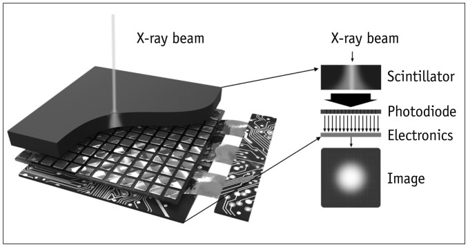
Fig. 1 Process of Gaussian modelling of light scattering in scintillator.
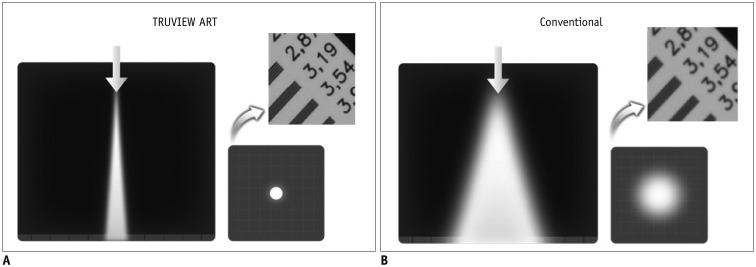
Fig. 2 Reconstructed image with and without TRUVIEW ART (DRTECH Corp.).A. By elimination of scattering effects applying TRUVIEW ART, blurred image can be seen more clearly. B. Without TRUVIEW ART, light scattering occurs by light spread of conventional scintillator, and image looks blurred.
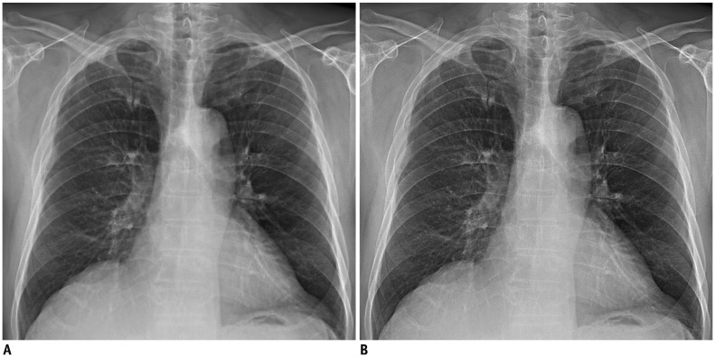
Fig. 3 75-year-old man with coronary artery disease.Compared with original chest radiography (A), TRUVEIW ART applied chest radiography (B) shows better depiction in overall image quality.

Fig. 4 61-year-old man with non-small cell lung cancer.Compared with original chest radiography (A, B), TRUVEIW ART applied chest radiography (C, D) shows better visualization of unobscured lung and thoracic spines.
Cited by 1 articles
-
The Potential Role of Grid-Like Software in Bedside Chest Radiography in Improving Image Quality and Dose Reduction: An Observer Preference Study
Su Yeon Ahn, Kum Ju Chae, Jin Mo Goo
Korean J Radiol. 2018;19(3):526-533. doi: 10.3348/kjr.2018.19.3.526.
Reference
-
1. Lee DL, Cheung LK, Rodricks BG, Powell GF. Improved imaging performance of a 14“×17” direct radiography system using a se/tft detector. Proc SPIE. 1998; 14–23.
Article2. Zhao W, Blevis I, Germann S, Rowlands JA, Waechter D, Huang Z. Digital radiology using active matrix readout of amorphous selenium: construction and evaluation of a prototype realtime detector. Med Phys. 1997; 24:1834–1843. PMID: 9434966.
Article3. Siewerdsen JH, Antonuk LE, el-Mohri Y, Yorkston J, Huang W, Boudry JM, et al. Empirical and theoretical investigation of the noise performance of indirect detection, active matrix flat-panel imagers (AMFPIs) for diagnostic radiology. Med Phys. 1997; 24:71–89. PMID: 9029542.
Article4. Colbeth RE, Cooper VN, Gilblom DL, Harris RA, Job ID, Klausmeier-Brown ME, et al. Characterization of a thirdgeneration multimode sensor panel. Proc SPIE. 1999; 491–500.5. Samei E, Flynn MJ. An experimental comparison of detector performance for direct and indirect digital radiography systems. Med Phys. 2003; 30:608–622. PMID: 12722813.
Article6. Fish DA, Brinicombe AM, Pike ER, Walker JG. Blind deconvolution by means of the Richardson-Lucy algorithm. J Opt Soc Am A. 1995; 12:58–65.
Article7. Levin A, Fergus R, Durand F, Freeman WT. Deconvolution using natural image priors. Massachusetts Institute of Technology, Computer Science and Artificial Intelligence Laboratory. 2007. 1. Accessed May 12, 2017. https://groups.csail.mit.edu/graphics/CodedAperture/SparseDeconv-LevinEtAl07.pdf.8. Peng-Xiang J, Feng Z, Bin Y, Shang-Lian B. Scattering correction method for panel detector based cone beam computed tomography system. Chinese Physics B. 2010; 19:087802.
Article
- Full Text Links
-
- Actions
-
Cited
- CITED
-
- Close
- Share
-
- Similar articles
-
- Erratum: Application of Deconvolution Algorithm of Point Spread Function in Improving Image Quality: An Observer Preference Study on Chest Radiography
- The Potential Role of Grid-Like Software in Bedside Chest Radiography in Improving Image Quality and Dose Reduction: An Observer Preference Study
- Objective and Quantitative Evaluation of Image Quality Using Fuzzy Integral: Phantom Study
- A New Full-Field Digital Mammography System with and without the Use of an Advanced Post-Processing Algorithm: Comparison of Image Quality and Diagnostic Performance
- Relationship of Image Quality and Radiation Dose for Chest Radiography in the Medical Institutions for Pneumoconiosis: A Comparison to the Korean Diagnostic Reference Level
