J Korean Soc Echocardiogr.
1994 Dec;2(2):155-163. 10.4250/jkse.1994.2.2.155.
The Parameters for the Evaluation of the Conduit Arterial Function Using Pulsed Doppler Echocardiography
- Affiliations
-
- 1Department of Internal Medicine, Wonkwang University School of Medicine, Iri, Korea.
- KMID: 2410443
- DOI: http://doi.org/10.4250/jkse.1994.2.2.155
Abstract
- BACKGROUND
Arterial change caused by of atherosclerosis in hypertensive or diabetic patients has been known to precede the symptoms resulting from occlusive disease of vital organs, such as heart, brain and kidney etc. The pulse-wave velocity of the pressure and flow waves produced during ventircular ejection has been shown to be a sensitive indicator of the physical properties of the arterial wall. Hence, measurements of pulse-wave velocith may be used to evaluate the arterial function.
OBJECTIVES
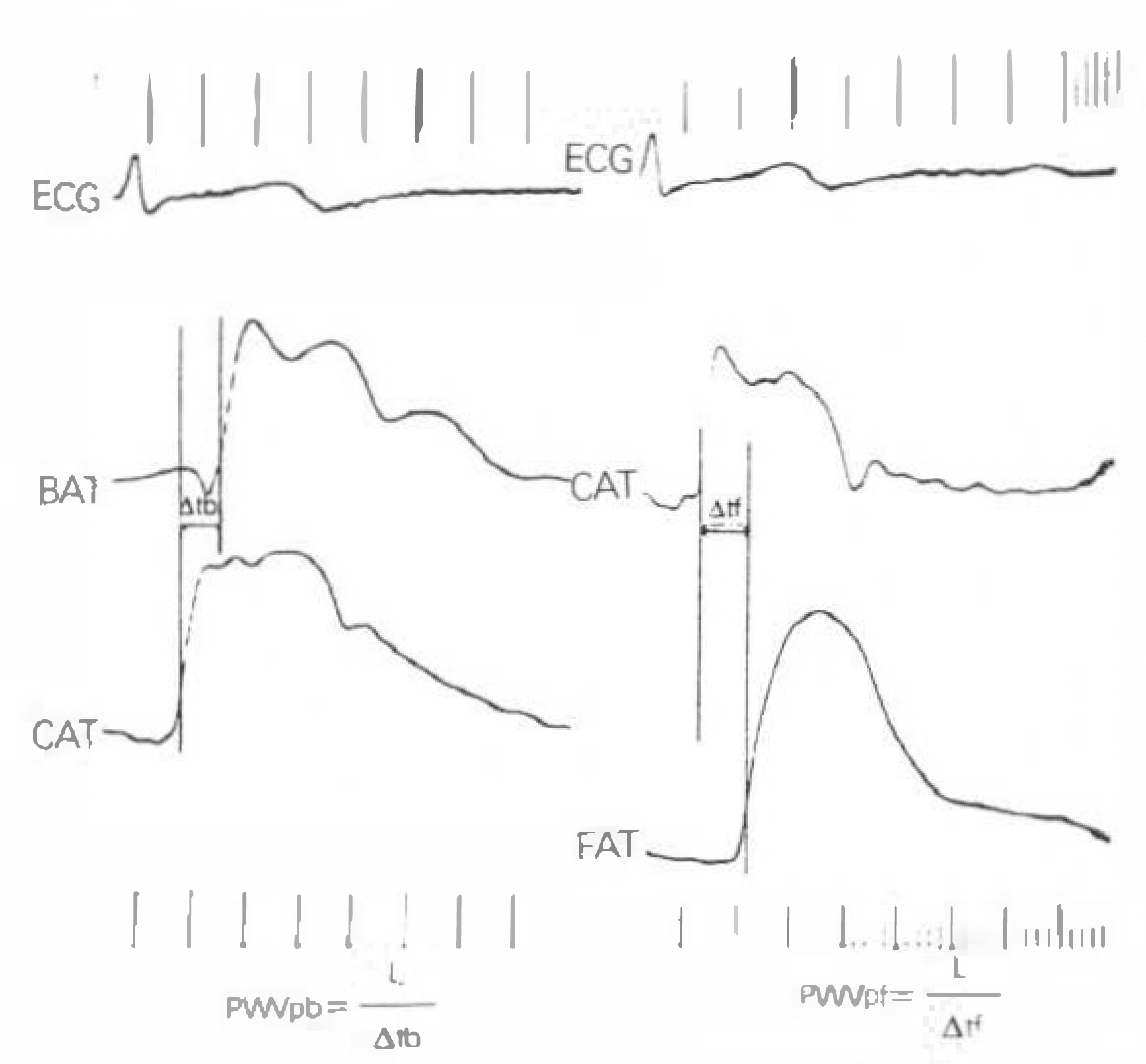
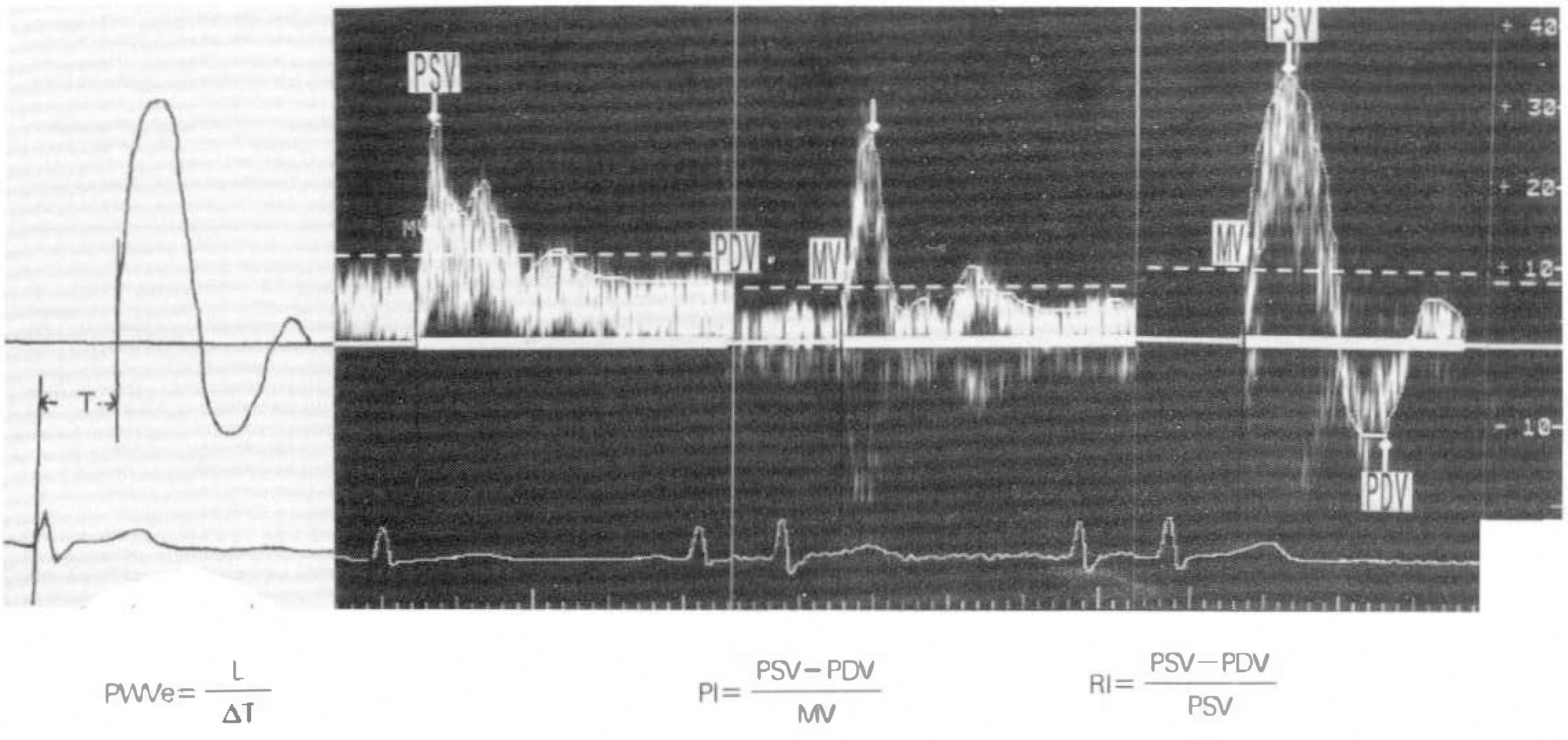
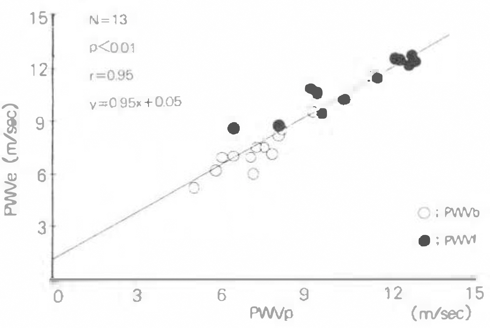
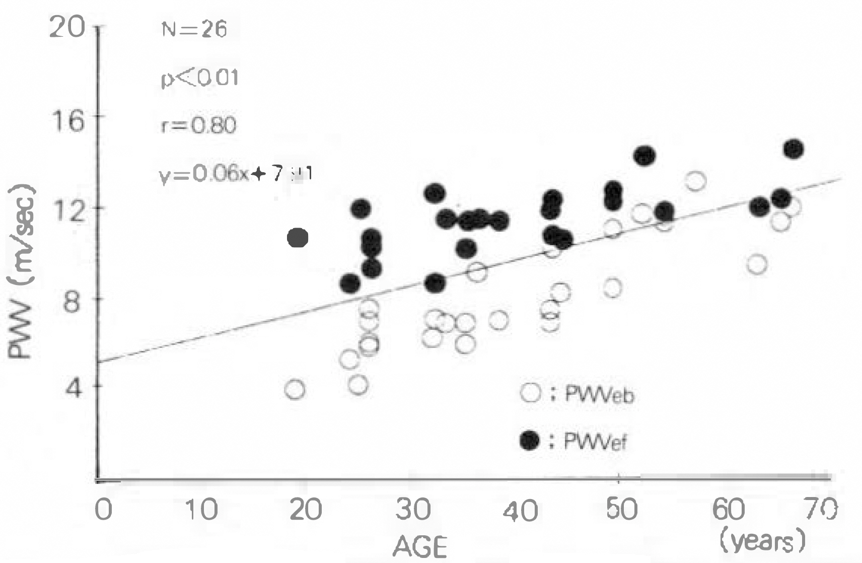
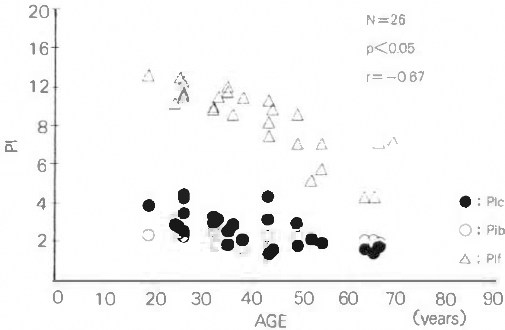
We performed this study to compare the pulse-wave velocity(PWV) measured by polygraph with that by the pulsed Doppler echocardiograph and to evaluate the correlation between Doppler echocardiographic pulsed-wave velocity, pulsatility index(PI) and resistive index(RI) and the age. METHOD: 26 normal subjects were screened for participation in this study to rule out any evidence of heart disease or peripheral vascular disease. PWV was measured in 16 subjects by polygraph(Honeywell VR6) and in all by pulse Doppler echocardiograph(ATL ultramark 9) at right common carotid artery, brachial artery and femoral artery with simultaneous recording of ECG(standard lead II). PI and RI were calculated by installed computer analyzer. RESULT: 1) There was no definite difference between PWV measured by polygraph and by pulse Doppler echocardiograph(74±1.6m/sec in upper and 10.6±2.0m/sec in lower extremity versus 7.6±1.6m/sec in upper extremity and 11.4+1.5m/sec in lower extremity). 2) PWV in both upper and lower extremities were influenced by the age of subjects(r=0.88, p < 0.01 and r=0.75, p < 0.01) and they were correlated significantly with each other(r=0.74, p < 0.01). 3) Both PI and Ri of right common carotid artery, brachial artery and femoral artery were correlated negatively with the age of subjects(r=−0.8, p < 0.01, r=−0.67, p < 0.01 and r=−0.84, p < 0.01 in PI and r=−0.66, p < 0.01, r=−0.58, p < 0.01 and r=−0.61, p < 0.01 in RI). 4) Significant negative correlations were also observed both between PWV or upper extremity and PI or RI in right common carotid artery and brachial artery(r=−0.71, p < 0.01 and r=−0.46, p < 0.05 in PI and r=−0.59, p < 0.01 and r=−0.56, p < 0.05 in RI) and between PWV of lower extremity and PI or RI in right common carotid artey and femoral artery(r=−0.65, p < 0.01 and r=0.53, p < 0.01 in PI and r=−0.54, p < 0.01 and r=−0.41, p < 0.05 in RI).
CONCLUSION
We conclude that PWV, PI and RI measured by pulsed Doppler echocardiograph is very useful in the evaluation of the function of conduit arteries.
MeSH Terms
Figure
Reference
-
References
1). Arnold JMO, Marchiori GE, Imrie JR, Burton GL, Pflugfelder PW, Kostuk JR. Large artery function in patient with chronic heart failure, Studies of brachial artery diameter and hemodynamics. Circulation. 94:2418. 1991.2). Cohn JN, Finkelstein SM. Abnormalities of vascular compliance in hypertension aging and heart failure. J Hypertension (Supp6). 61:1992.
Article3). Gribbin B, Stepto A, Sleighr P. Pulse wave velocity as a measure of blood pressure change. Psychophysiology. 13:86. 1976.
Article4). Rifkin MD, Needleman L, Pasto O, Kurtz AB, Foy PM, McGlynn E, Canino C, Blatarocoich OH, Pennel RG, Goldberg BB. Evaluation of renal transplant rejection by Duplex Doppler examination: Value of the resistive index. Am J Radiol. 148:759. 1987.
Article5). Stuat B, Dramm J, Fitzgerald DE, Duigan NM. Fetal blood velocity waveforms in normal pregnancy. Br J Obstet Gynaecol. 87:780. 1980.6). Gosing RG, King DH. Ultrasound angiology in arteries and veins. Marcus AW, Adamson L, editors. Churchill Livingstone Edinburgh;p. 61–98. 1975.7). 이 좋 녁 • 이 지 영 • 김 창단 • 정 선 판 · 노 병 씌 • 왼 종 진 · 정l홉· 분딴현탁액 주입후 가토 대퇴동맥에서의 힌류속도파형 변화에 대한 실험 적 연구. 대한초유 파의학회 지. 2:152. 1990.8). Legarth J, Thorup E. Characteristics of Doppler blood-velocity waveforms in a cardiovascular in vitro model II. The influence peripheral resistance, perfusion pressure and blood flow. Scand J Clin Lab Invest. 89:451. 1989.9). Wezler K. Die Anwedung der physikalischen Methoden der Schlag volumbestimmung. Verh, dutsch, Ges, Kleisl, Forsch. 15(Anhang):18. 1949.10). 흡題 ·μ格훤義明·字흉뿜文村物 的心服·환맹的 分析法呼. 12:15. 1964.11). 朴E후, 鄭泰핸 • 朴良主; 韓國正1f; A에 있어서縮빠間間隔 非觀血的血力學態및 心賢收 縮의릅標에 關한�. 순환기. 9:1. 1979.12). Scarpello JHB, Martin TRP, Ward JD. Ultrasound measurements of pulse wave velocity in the peripheral wave velocity in the peripheral arteries of diabetic subjects. Clin Science. 58:53. 1980.13). Simon AC, Levenson J, Bouthier J, Sater ME, Avolo AP. Evidence of early degenerative changes in large arteres in human essential hypertension. Hypertension. 7:675. 1985.14). O'Rourke M. Arterial Stiffness, Systolic blood pressure and logical treatment of arterial hypertension. Hypertension. 15:339. 1990.15). Isard RN, Pannier BM, Laurent S, London GM, Diebold B, Safar ME. Pulsatile diamenter and elastic modulus of the aortic arch in essential hypertension. A noninvasive study. J Am Coll Cardiol. 13:399. 1989.16). Edmonds ME, Roberts VC, Watkins PJ. Blood flow in the Diabetic Neuropathic foot. Diabetolgia. 22:9. 1982.
Article17). Kelly RP, Gibbs HU, O'Rourke MF, Daley JE, Mang K, Morgan JJ, Avolio AP. Nitroglycerin has more favourable effects on left ventricular afterload than apparent from measurement of pressure in a peripheral artery. Eur Heart J. 11:138. 1990.
Article18). Hirai T, Sasyama S, Kwasaki T, Yagi S. Stiffness of systemic arteries in patients with myocardial infarction. A noninvasive method to predict severity of coronary artherosclerosis. Circulation. 80:78. 1989.19). Brawell JV, Hill AV. Velocity of transmission of the pulse wave and elasticity of arteries. Lancet. 1:891. 1922.20). Avolio AP, Denz FQ, Luo YF, Huang ZD, Xing LF, O'Rourke MF. Effects of aging on arterial distensibility in populations with high and low prevalence of hypertension comparison between rual communities in china. Circulation. 71:202. 1985.21). Kawasaki T, Sasayama S, Yagi S, Asakawa T, Hirai T. Nonivasive assessment of the age related change in stiffness of major branches of the human arteries. Cardiovase Res. 21:678. 1987.22). Woolam GL, Schnur PL, Vallbona C, Hoft HE. The Pulse wave velocity as an early indicator of atherosclerosis in diabetice subjects. Circulation. 25:533. 1962.23). Schulman H. The clinical implications of Doppler ultrasound analysis of the uterine and umbilical arteries. Am J Obstet Gynecol. 156:889. 1987.
Article24). Griffin D, Bilardo K, Mashini L, Diazrecasens J, Perace JM, Willson K. Campbells, Doppler blood flow waveforms in the descending thoracic aorta of the human fetus. British J Obstetrics Gynecology. 91:997. 1984.25). Mires G, Dempster J, Patel NB, Crawford JW. The effect of the fetal heart rate on umbilical artery flow velocity waveforms. British J Obstetrics Gynecology. 94:665. 1987.26). Daprez DA, De Buyzere ML, Debacker T, Kaufman JM, Van Hoeck MJ, Vermealen A, Coment DL. Influence of systemic arterial blood pressure and nonhemodynamic factors on the brachial arterial pulsatility index in mild to moderate essential hypertension. Am J Cardiol. 71:350. 1993.27). Yoshimura S, Kodaira K, Fujishiro K, Furuhata H. A newly developed noninvasive technique for quantitative measurement of blood flow with special reference to the measurement of carotid arterial blood flow. Jikeikai Med J. 28:241. 1981.28). Duprez D, De Buyzere M, Brusselmans F, Meas A, Clement DL. Comparsion of lisinopril and nitrendipine on the pulsatility index in mild essential arterial hypertension. Cardiovascular Drugs and Therapy. 6:399. 1992.
- Full Text Links
- Actions
-
Cited
- CITED
-
- Close
- Share
- Similar articles
-
- Value of Pulsed Doppler Echocardiography in the Diagnosis of Aortic Regurgitation
- Pulsed doppler echocardiographic analysis of pulmonary venous flow in congenital heart diseases with left-to-right shunt
- Doppler ultrasonography of the lower extremity arteries: anatomy and scanning guidelines
- Study of left and right ventricular diastolic dysfunction in the hypertensive patients by pulsed doppler echocardiography
- Reconstruction of the Transmitral Flow Rate Curve with M-Mode,2-Dimensional and Doppler Echocardiography -Validation Study-

