J Adv Prosthodont.
2018 Apr;10(2):163-166. 10.4047/jap.2018.10.2.163.
Clinical outcome of immediately and early loaded implants with laser treated surface: a 3-year retrospective study
- Affiliations
-
- 1Department of Biomaterials & Prosthodontics, Kyung Hee University Hospital at Gangdong, School of Dentistry, Kyung Hee University, Seoul, Republic of Korea.
- 2Department of Prosthodontics, School of Dentistry, Kyung-Pook National University, Daegu, Republic of Korea. sungamcho@gamil.com
- KMID: 2409637
- DOI: http://doi.org/10.4047/jap.2018.10.2.163
Abstract
- PURPOSE
The marginal bone loss of implants with laser treated surface was investigated after six weeks of loading after implant installation to the mandible molar area.
MATERIALS AND METHODS
A total of 23 implants were placed in the edentulous molar area of the mandible: 13 implants were immediately loaded and 10 implants were early loaded. The implants used were made of titanium grade 23, screw shaped, 4.2 mm in diameter, and 10 mm in length. Patients were evaluated with resonance frequency analysis at implant fixture installation and 1, 2 (final prosthesis installation), 3, 5, 8, and 14 months later. X-rays were taken at 2 months after fixture installation and 1, 2, 3 years after to measure the marginal bone loss.
RESULTS
The mean ISQ value measured at the implant installation was over 70 at all-time points. The average of marginal bone loss was average 0.33 mm.
CONCLUSION
Immediate implant loading for laser treated implants would be possible.
Figure
-
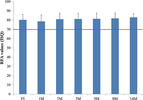
Fig. 1 Mean and SD of ISQ values measured from implant fixture installation to 14 months after surgery. FI: at implant installation, 1M: 1 month later, 2M: 2 months later (final prosthesis placement), 3M: 3 months later, 5M: 5 months later, 8M: 8 months later, 14M: 14 months later. Success criterion was a value greater than or equal to 70 (red-line).
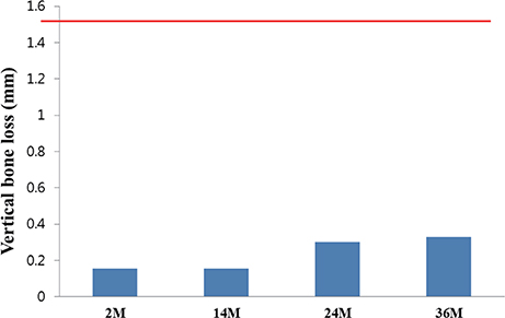
Fig. 2 Mean and SD of marginal bone loss measured from implant fixture installation to 36 months after surgery. 2M: 2 months later (final prosthesis placement), 14M: 14 months later, 24M: 24 months later, 36M: 36 months later. Cut-off value was 1.5 mm (red-line).
Reference
-
1. Wismeijer D, Casentini P, Gallucci G, Chiapasco M. ITI treatment guide Volume 4, Loading protocol in implant dentistry. Quintessence;2010. p. 6–8.2. Rupp F, Scheideler L, Olshanska N, de Wild M, Wieland M, Geis-Gerstorfer J. Enhancing surface free energy and hydrophilicity through chemical modification of microstructured titanium implant surfaces. J Biomed Mater Res A. 2006; 76:323–334.
Article3. Hofmann AA, Bloebaum RD, Bachus KN. Progression of human bone ingrowth into porous-coated implants. Rate of bone ingrowth in humans. Acta Orthop Scand. 1997; 68:161–166.
Article4. Zhao G, Schwartz Z, Wieland M, Rupp F, Geis-Gerstorfer J, Cochran DL, Boyan BD. High surface energy enhances cell response to titanium substrate microstructure. J Biomed Mater Res A. 2005; 74:49–58.
Article5. Wennerberg A, Galli S, Albrektsson T. Current knowledge about the hydrophilic and nanostructured SLActive surface. Clin Cosmet Investig Dent. 2011; 3:59–67.
Article6. Hall J, Miranda-Burgos P, Sennerby L. Stimulation of directed bone growth at oxidized titanium implants by macroscopic grooves: an in vivo study. Clin Implant Dent Relat Res. 2005; 7:S76–S82.
Article7. Mangano C, Perrotti V, Iezzi G, Scarano A, Mangano F, Piattelli A. Bone response to modified titanium surface implants in nonhuman primates (Papio ursinus) and humans: histological evaluation. J Oral Implantol. 2008; 34:17–24.8. Schwartz Z, Martin JY, Dean DD, Simpson J, Cochran DL, Boyan BD. Effect of titanium surface roughness on chondrocyte proliferation, matrix production, and differentiation depends on the state of cell maturation. J Biomed Mater Res. 1996; 30:145–155.
Article9. Roccuzzo M, Bunino M, Prioglio F, Bianchi SD. Early loading of sandblasted and acid-etched (SLA) implants: a prospective split-mouth comparative study. Clin Oral Implants Res. 2001; 12:572–578.
Article10. Cochran DL, Buser D, ten Bruggenkate CM, Weingart D, Taylor TM, Bernard JP, Peters F, Simpson JP. The use of reduced healing times on ITI implants with a sandblasted and acid-etched (SLA) surface: early results from clinical trials on ITI SLA implants. Clin Oral Implants Res. 2002; 13:144–153.11. Cochran D, Oates T, Morton D, Jones A, Buser D, Peters F. Clinical field trial examining an implant with a sand-blasted, acid-etched surface. J Periodontol. 2007; 78:974–982.
Article12. Hallgren C, Reimers H, Chakarov D, Gold J, Wennerberg A. An in vivo study of bone response to implants topographically modified by laser micromachining. Biomaterials. 2003; 24:701–710.
Article13. Kang NS, Li LJ, Cho SA. Comparison of removal torques between laser-treated and SLA-treated implant surfaces in rabbit tibiae. J Adv Prosthodont. 2014; 6:302–308.
Article14. Gaggl A, Schultes G, Müller WD, Kärcher H. Scanning electron microscopical analysis of laser-treated titanium implant surfaces-a comparative study. Biomaterials. 2000; 21:1067–1073.
Article15. Cho SA, Jung SK. A removal torque of the laser-treated titanium implants in rabbit tibia. Biomaterials. 2003; 24:4859–4863.
Article
- Full Text Links
-
- Actions
-
Cited
- CITED
-
- Close
- Share
-
- Similar articles
-
- Erratum - Clinical outcome of immediately and early loaded implants with laser treated surface: a 3-year retrospective study
- Marginal bone level change during sequential loading periods of partial edentulous rehabilitation using immediately loaded selftapping implants: a 6.5-year retrospective study
- The effects of saline soaking on the removal torque of titanium implants in rabbit tibia after 10 days
- The effect of early loading on the direct bone-to-implant surface contact of the orthodontic osseointegrated titanium implant
- Fatigue resistance of dental implants treated with laser method
