Investig Magn Reson Imaging.
2018 Mar;22(1):56-60. 10.13104/imri.2018.22.1.56.
Complete Recovery of Perfusion Abnormalities in a Cardiac Arrest Patient Treated with Hypothermia: Results of Cerebral Perfusion MR Imaging
- Affiliations
-
- 1Department of Radiology, Ewha Womans University College of Medicine, Seoul, Korea. yaewonpark@yuhs.ac
- KMID: 2408818
- DOI: http://doi.org/10.13104/imri.2018.22.1.56
Abstract
- Therapeutic hypothermia in cardiac arrest patients is associated with favorable outcomes mediated via neuroprotective mechanisms. We report a rare case of a 32-year-old male who demonstrated complete recovery of signal changes on perfusion-weighted imaging after therapeutic hypothermia due to cardiac arrest. Brain MRI with perfusion-weighted imaging, performed three days after ending the hypothermia therapy, showed a marked decrease in relative cerebral blood flow (rCBF) and delay in mean transit time (MTT) in the bilateral basal ganglia, thalami, brain stem, cerebellum, occipitoparietal cortex, and frontotemporal cortex. However, no cerebral ischemia was not noted on diffusion-weighted imaging (DWI) or fluid-attenuated inversion recovery (FLAIR) sequences. A follow-up brain MRI after one week showed complete resolution of the perfusion deficit and the patient was discharged without any neurologic sequelae. The mechanism and interpretation of the perfusion changes in cardiac arrest patients treated with therapeutic hypothermia are discussed.
Keyword
MeSH Terms
Figure
-
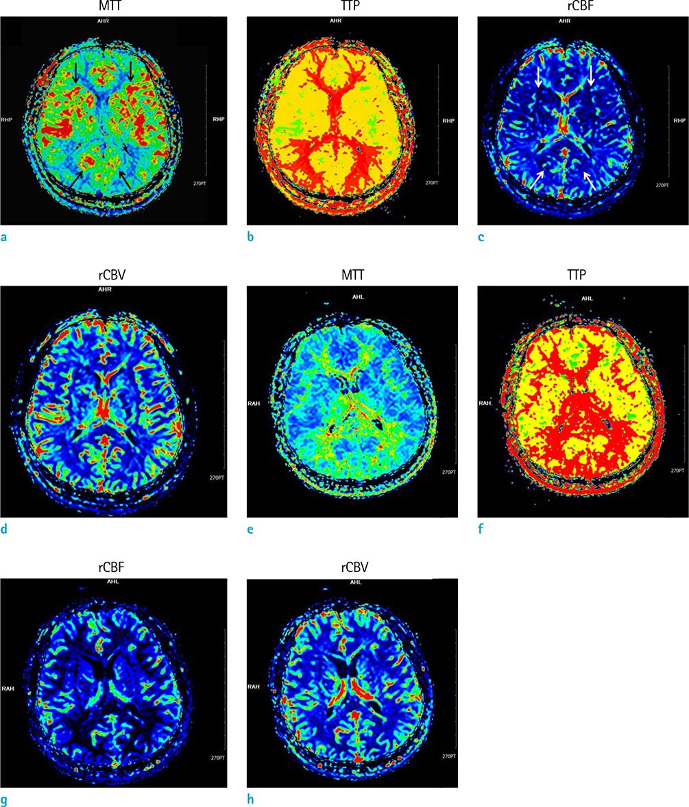
Fig. 1 Initial brain perfusion-weighted imaging (a-d) showed delay in mean transit time (MTT) with decreased relative cerebral blood flow (rCBF) in bilateral basal ganglia, thalami, occipital cortex and temporal cortex (arrows in a, c), which were recovered on follow-up imaging (e-h). rCBV = relative cerebral blood volume; TTP = time to peak
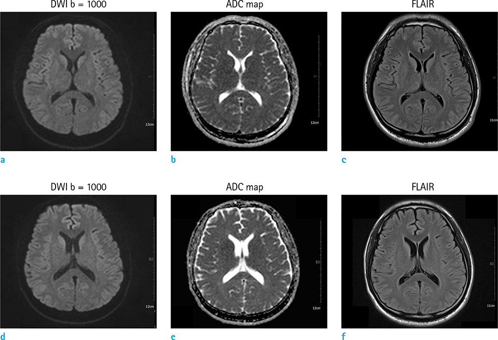
Fig. 2 Diffusion-weighted imaging (DWI), apparent diffusion coefficient (ADC) map, and fluid-attenuated inversion recovery (FLAIR) imaging showed no evidence of ischemic changes in initial (a-c) and follow-up (d-f) imaging.
Reference
-
1. Holzer M, Bernard SA, Hachimi-Idrissi S, et al. Hypothermia for neuroprotection after cardiac arrest: systematic review and individual patient data meta-analysis. Crit Care Med. 2005; 33:414–418.
Article2. Li F, Liu KF, Silva MD, et al. Acute postischemic renormalization of the apparent diffusion coefficient of water is not associated with reversal of astrocytic swelling and neuronal shrinkage in rats. AJNR Am J Neuroradiol. 2002; 23:180–188.3. Jarnum H, Knutsson L, Rundgren M, et al. Diffusion and perfusion MRI of the brain in comatose patients treated with mild hypothermia after cardiac arrest: a prospective observational study. Resuscitation. 2009; 80:425–430.
Article4. Liu R, Li X, Hu CL, et al. The changes of brain water diffusion and blood flow on diffusion-weighted and perfusion-weighted imaging in a canine model of cardiac arrest. Resuscitation. 2012; 83:645–651.
Article5. Youn CS, Park KN, Kim JY, et al. Repeated diffusion weighted imaging in comatose cardiac arrest patients with therapeutic hypothermia. Resuscitation. 2015; 96:1–8.
Article6. Keller E, Steiner T, Fandino J, Schwab S, Hacke W. Changes in cerebral blood flow and oxygen metabolism during moderate hypothermia in patients with severe middle cerebral artery infarction. Neurosurg Focus. 2000; 8:e4.
Article7. Huang BY, Castillo M. Hypoxic-ischemic brain injury: imaging findings from birth to adulthood. Radiographics. 2008; 28:417–439. quiz 617.
Article
- Full Text Links
-
- Actions
-
Cited
- CITED
-
- Close
- Share
-
- Similar articles
-
- Cerebral Oxygen Saturation Monitoring during Aortic Dissection Surgery: A case report
- Selective Cerebral Perfusion Through the Right Axillary Artery for Aortic Arch Replacement: A Case Report
- Anesthetic Management of Patient with Renal Cell Carcinoma Extending into the Right Atrium Using Adjunctive Deep Hypothermic Circulatory Arrest: A case report
- Repair of Acute Aortic Arch Dissection with Hypothermic Circulatory Arrest and Retrograde Cerebral Perfusion
- Analysis of Neurological Complications on Antegrade Versus Retrograde Cerebral Perfusion in the Surgical Treatment of Aortic Dissection
