J Vet Sci.
2018 Mar;19(2):200-206. 10.4142/jvs.2018.19.2.200.
Packaging of Rift Valley fever virus pseudoviruses and establishment of a neutralization assay method
- Affiliations
-
- 1College of Animal Science and Veterinary Medicine, Henan Institute of Science and Technology, Xinxiang 453003, China. xiaxzh@cae.cn
- 2Institute of Military Veterinary Medicine, Academy of Military Medical Science, Changchun 130122, China.
- 3College of Animal Science and Technology, Shihezi University, Shihezi 832000, China.
- 4School of Public Health, Shandong University, Jinan 250100, China.
- 5College of Veterinary Medicine, Jilin University, Changchun 130012, China.
- 6Changchun SR Biological Technology Co., Ltd., Changchun 130000, China.
- KMID: 2407619
- DOI: http://doi.org/10.4142/jvs.2018.19.2.200
Abstract
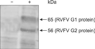
- Rift Valley fever (RVF) is an acute, febrile zoonotic disease that is caused by the RVF virus (RVFV). RVF is mainly prevalent on the Arabian Peninsula, the African continent, and several islands in the Indian Ocean near southeast Africa. RVFV has been classified by the World Organisation for Animal Health (OIE) as a category A pathogen. To avoid biological safety concerns associated with use of the pathogen in RVFV neutralization assays, the present study investigated and established an RVFV pseudovirus-based neutralization assay. This study used the human immunodeficiency virus (HIV) lentiviral packaging system and RVFV structural proteins to successfully construct RVFV pseudoviruses. Electron microscopy observation and western blotting indicated that the size, structure, and shape of the packaged pseudoviruses were notably similar to those of HIV lentiviral vectors. Infection inhibition assay results showed that an antibody against RVFV inhibited the infective ability of the RVFV pseudoviruses, and an antibody neutralization assay for RVFV detection was then established. This study has successfully established a neutralization assay based on RVFV pseudoviruses and demonstrated that this method can be used to effectively evaluate antibody neutralization.
MeSH Terms
Figure
Reference
-
1. Anderson GW Jr, Slone TW Jr, Peters CJ. The gerbil, Meriones unguiculatus, a model for Rift Valley fever viral encephalitis. Arch Virol. 1988; 102:187–196.
Article2. Ashrafi GH, Piuko K, Burden F, Yuan Z, Gault EA, Müller M, Trawford A, Reid SW, Nasir L, Campo MS. Vaccination of sarcoid-bearing donkeys with chimeric virus-like particles of bovine papillomavirus type 1. J Gen Virol. 2008; 89:148–157.
Article3. Bartosch B, Bukh J, Meunier JC, Granier C, Engle RE, Blackwelder WC, Emerson SU, Cosset FL, Purcell RH. In vitro assay for neutralizing antibody to hepatitis C virus: evidence for broadly conserved neutralization epitopes. Proc Natl Acad Sci U S A. 2003; 100:14199–14204.
Article4. Bartosch B, Dubuisson J, Cosset FL. Infectious hepatitis C virus pseudo-particles containing functional E1-E2 envelope protein complexes. J Exp Med. 2003; 197:633–642.
Article5. Boshra H, Lorenzo G, Busquets N, Brun A. Rift valley fever: recent insights into pathogenesis and prevention. J Virol. 2011; 85:6098–6105.
Article6. Brown BD, Sitia G, Annoni A, Hauben E, Sergi LS, Zingale A, Roncarolo MG, Guidotti LG, Naldini L. In vivo administration of lentiviral vectors triggers a type I interferon response that restricts hepatocyte gene transfer and promotes vector clearance. Blood. 2007; 109:2797–2805.
Article7. Giroglou T, Cinatl J Jr, Rabenau H, Drosten C, Schwalbe H, Doerr HW, von Laer D. Retroviral vectors pseudotyped with severe acute respiratory syndrome coronavirus S protein. J Virol. 2004; 78:9007–9015.
Article8. Hilgenfeld R, Peiris M. From SARS to MERS: 10 years of research on highly pathogenic human coronaviruses. Antiviral Res. 2013; 100:286–295.
Article9. Hofmann H, Hattermann K, Marzi A, Gramberg T, Geier M, Krumbiegel M, Kuate S, Uberla K, Niedrig M, Pöhlmann S. S protein of severe acute respiratory syndrome-associated coronavirus mediates entry into hepatoma cell lines and is targeted by neutralizing antibodies in infected patients. J Virol. 2004; 78:6134–6142.
Article10. Ikegami T. Molecular biology and genetic diversity of Rift Valley fever virus. Antiviral Res. 2012; 95:293–310.
Article11. Ikegami T, Makino S. The pathogenesis of Rift Valley fever. Viruses. 2011; 3:493–519.
Article12. Kim YB, Lee MK, Han DP, Cho MW. Development of a safe and rapid neutralization assay using murine leukemia virus pseudotyped with HIV type 1 envelope glycoprotein lacking the cytoplasmic domain. AIDS Res Hum Retroviruses. 2001; 17:1715–1724.
Article13. Li C, Bu Z, Chen H. Avian influenza vaccines against H5N1 ‘bird flu’. Trends Biotechnol. 2014; 32:147–156.
Article14. Li M, Gao F, Mascola JR, Stamatatos L, Polonis VR, Koutsoukos M, Voss G, Goepfert P, Gilbert P, Greene KM, Bilska M, Kothe DL, Salazar-Gonzalez JF, Wei X, Decker JM, Hahn BH, Montefiori DC. Human immunodeficiency virus type 1 env clones from acute and early subtype B infections for standardized assessments of vaccine-elicited neutralizing antibodies. J Virol. 2005; 79:10108–10125.
Article15. Nie Y, Wang G, Shi X, Zhang H, Qiu Y, He Z, Wang W, Lian G, Yin X, Du L, Ren L, Wang J, He X, Li T, Deng H, Ding M. Neutralizing antibodies in patients with severe acute respiratory syndrome-associated coronavirus infection. J Infect Dis. 2004; 190:1119–1126.16. Niklasson B, Peters CJ, Grandien M, Wood O. Detection of human immunoglobulins G and M antibodies to Rift Valley fever virus by enzyme-linked immunosorbent assay. J Clin Microbiol. 1984; 19:225–229.
Article17. Paweska JT, Burt FJ, Anthony F, Smith SJ, Grobbelaar AA, Croft JE, Ksiazek TG, Swanepoel R. IgG-sandwich and IgM-capture enzyme-linked immunosorbent assay for the detection of antibody to Rift Valley fever virus in domestic ruminants. J Virol Methods. 2003; 113:103–112.
Article18. Qiu C, Huang Y, Zhang A, Tian D, Wan Y, Zhang X, Zhang W, Zhang Z, Yuan Z, Hu Y, Zhang X, Xu J. Safe pseudovirus-based assay for neutralization antibodies against influenza A(H7N9) virus. Emerg Infect Dis. 2013; 19:1685–1687.
Article19. Rivera A, Messaoudi I. Molecular mechanisms of Ebola pathogenesis. J Leukoc Biol. 2016; 100:889–904.
Article20. Simmons G, Reeves JD, Rennekamp AJ, Amberg SM, Piefer AJ, Bates P. Characterization of severe acute respiratory syndrome-associated coronavirus (SARS-CoV) spike glycoprotein-mediated viral entry. Proc Natl Acad Sci U S A. 2004; 101:4240–4245.
Article
- Full Text Links
- Actions
-
Cited
- CITED
-
- Close
- Share
- Similar articles
-
- A serological study of severe fever with thrombocytopenia syndrome using a virus neutralization test and competitive enzyme-linked immunosorbent assay
- SARS-CoV-2 Antibody Neutralization Assay Platforms Based on Epitopes Sources: Live Virus, Pseudovirus, and Recombinant S Glycoprotein RBD
- Development of Neutralization Assay using Murine Leukemia Virus (MuLV) Pseudotyped with Japanese encephalitis Virus (JEV) env Gene
- Identification of Retroviral Vectors Producing High Viral Titer
- Sero-survey on Aino, Akabane, Chuzan, bovine ephemeral fever and Japanese encephalitis virus of cattle and swine in Korea


