The Normal Aging Chest: An Overview of Radiologic Imaging Features
- Affiliations
-
- 1Department of Radiology, Korea University Guro Hospital, College of Medicine, Korea University, Seoul, Korea. keyrad@korea.ac.kr
- 2Department of Radiology, Korea University Ansan Hospital, College of Medicine, Korea University, Ansan, Korea.
- 3Department of Radiology, Korea University Anam Hospital, College of Medicine, Korea University, Seoul, Korea.
- KMID: 2405731
- DOI: http://doi.org/10.3348/jksr.2017.77.3.166
Abstract
- Many age-related morphologic changes are revealed in radiologic images of the chest in the elderly. We categorize the aging chest according to changes in the lung, airways, mediastinum, chest wall, and diaphragm. Changes in the lung include age-related alveolar hyperinflation, ground-glass opacity in basal dependent lungs, mosaic attenuation pattern, reticular densities in basal subpleural lungs, small nodule, air cyst, and apical cap. Changes in the airway include the tracheobronchial wall cartilage calcification, increased anterior-posterior diameter of the trachea, increased bronchoarterial ratio and bronchial wall thickness. Mediastinum changes include cardiac enlargement, coronary and cardiac valve/annulus calcification, aorta dilation and wall calcification, and excessive fat deposition. The chest wall shows decreased muscle mass, osteophytes, rib cartilage calcifications, and increased thoracic anterior-posterior diameter. The diaphragm changes include bulging contour, diaphragm defect, and esophageal hiatal hernia. Radiologists should therefore be aware of the age-related changes in the elderly chest. Differentiation between normal age-related changes and clinically significant disease is essential in the interpretation of chest radiologic images.
MeSH Terms
Figure
-
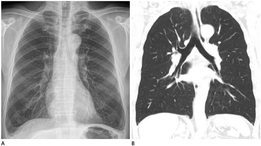
Fig. 1 Age-related alveolar hyperinflation in a 69-year-old man non-smoker. Chest radiograph (A) shows uniform hyperlucent lungs with flattened diaphragms, which mimic the findings of emphysema. However, chest CT (B) shows no emphysema.
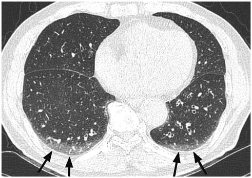
Fig. 2 Homogeneous ground glass opacity in the basal dependent lungs (arrows) seen on CT scan of a 69-year-old man. It is due to shallow inspiration depth and dependent position of the lung parenchyma, and is reversible in prone position.
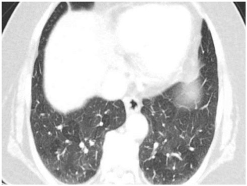
Fig. 3 Mosaic attenuation pattern in lower lungs in CT scan of a 76-year-old woman. It is caused by expiration or limited respiration during CT examination.

Fig. 4 Subtle reticular densities in both lower subpleural lungs on CT in an asymptomatic 76-year-old woman non-smoker. This limited subpleural basal reticular pattern is not associated with traction bronchiectasis or honeycombing.

Fig. 5 Focal reticular densities (arrow) in the right lower lobe, adjacent to a thoracic spine osteophyte, seen on CT in a 67-year-old woman.
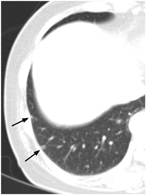
Fig. 6 Small nodules on the right lower lobe subpleural lung (arrows) on CT in a 55-year-old woman with uterine cervical cancer. These nodules were histologically confirmed as intrapulmonary lymph nodes. It should be remembered that subpleural nodules located in the lower lungs may be intrapulmonary lymph nodes, even though the patient has malignancy.

Fig. 7 A small ovoid nodule in the right major fissure (arrow), seen in CT of a 66-year-old woman. This perifissural nodule is consistent with intrapulmonary lymph node, and further follow-up is not required.

Fig. 8 CT showing a small thin-walled air cyst in the right middle lobe(arrow), in a 73-year-old woman.
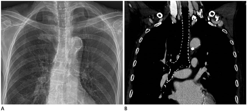
Fig. 9 Chest radiograph (A) and CT (B) show diffuse cartilage calcification along the tracheobronchial wall in 83-year-old-woman.
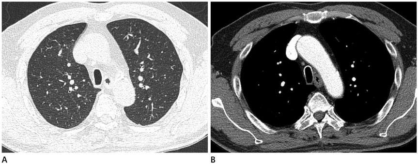
Fig. 10 Increased anterior-posterior diameter of intrathoracic trachea with diffuse tracheal wall calcification on CT (A, B) in a 74-year-old man. He is a past smoker, with no history of emphysema or chronic obstructive lung disease.
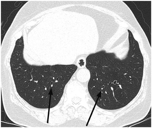
Fig. 11 Increased bronchoarterial ratio (arrows) in lower lungs on thin-section CT in asymptomatic 76-year-old woman. She is a non-smoker, and has no known lung disease.
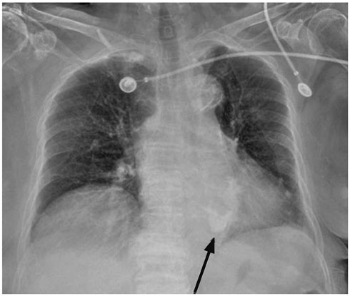
Fig. 12 Chest radiograph shows mild cardiomegaly, mitral annular calcification (arrow), and diffuse aortic wall calcification in a 92-year-old woman.
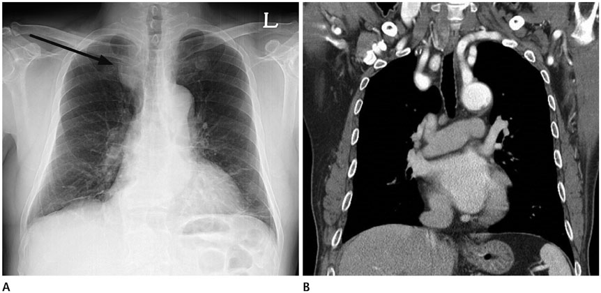
Fig. 13 Chest radiograph (A) in a 71-year-old man, shows a bulging soft tissue opacity in the right paratracheal area (arrow), corresponding to a tortuous right brachiocephalic to subclavian artery seen on CT (B).
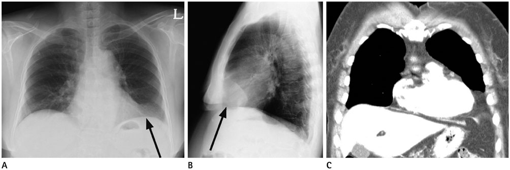
Fig. 14 Chest radiographs (A, B) in a 72-year-old woman show mass opacity (arrows) in the left cardiophrenic angle area, which is classic appearance of prominent paracardaic fat pad. This is confirmed on CT (C).
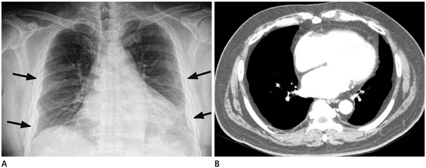
Fig. 15 Chest radiograph (A) in a 59-year-old man shows prominent epipleural fat (arrows) mimicking pleural thickening in both hemithoraces. This is confirmed by CT (B).
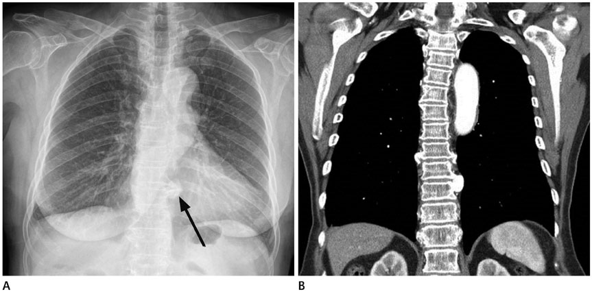
Fig. 16 Chest radiograph (A) in a 75-year-old woman shows a nodular opacity in the left paraspinal area (arrow), which corresponds to the bridging osteophytes on CT (B).
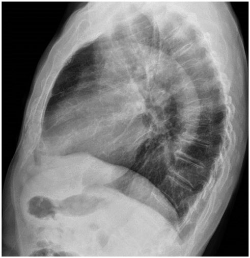
Fig. 17 Chest lateral radiograph shows increased thoracic anterior-posterior diameter in an asymptomatic 74-year-old man. He has no emphysema and chronic obstructive lung disease.
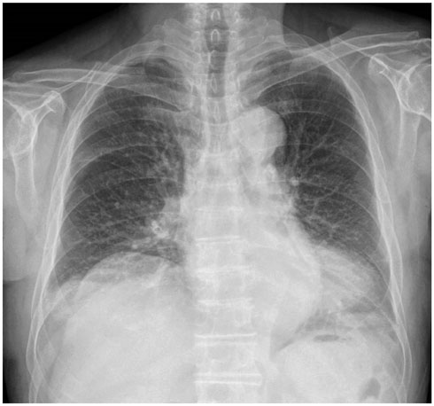
Fig. 18 Chest radiograph shows elevation and bulging contour in right diaphragm in an 84-year-old man.

Fig. 19 Chest radiograph (A) in an 80-year-old woman shows a large mass opacity with internal air density in the left retrocardiac area (arrows). Chest CT (B) confirms esophageal hiatal hernia. Increased anterior-posterior distance of the thorax causes stretching of the diaphragm, resulting in widening of the esophageal hiatus.
Reference
-
1. Miller MR. Structural and physiological age-associated changes in aging lungs. Semin Respir Crit Care Med. 2010; 31:521–527.2. Schröder TH, Storbeck B, Rabe KF, Weber C. The aging lung: clinical and imaging findings and the fringe of physiological state. Rofo. 2015; 187:430–439.3. The definition of emphysema. Report of a National Heart, Lung, and Blood Institute, Division of Lung Diseases workshop. Am Rev Respir Dis. 1985; 132:182–185.4. Well DS, Meier JM, Mahne A, Houseni M, Hernandez-Pampaloni M, Mong A, et al. Detection of age-related changes in thoracic structure and function by computed tomography, magnetic resonance imaging, and positron emission tomography. Semin Nucl Med. 2007; 37:103–119.5. Copley SJ, Giannarou S, Schmid VJ, Hansell DM, Wells AU, Yang GZ. Effect of aging on lung structure in vivo: assessment with densitometric and fractal analysis of high-resolution computed tomography data. J Thorac Imaging. 2012; 27:366–371.6. Lee KW, Chung SY, Yang I, Lee Y, Ko EY, Park MJ. Correlation of aging and smoking with air trapping at thin-section CT of the lung in asymptomatic subjects. Radiology. 2000; 214:831–836.7. Copley SJ, Wells AU, Hawtin KE, Gibson DJ, Hodson JM, Jacques AE, et al. Lung morphology in the elderly: comparative CT study of subjects over 75 years old versus those under 55 years old. Radiology. 2009; 251:566–573.8. Calabresi C, Arosio B, Galimberti L, Scanziani E, Bergottini R, Annoni G, et al. Natural aging, expression of fibrosis-related genes and collagen deposition in rat lung. Exp Gerontol. 2007; 42:1003–1011.9. Bonomo L, Larici AR, Maggi F, Schiavon F, Berletti R. Aging and the respiratory system. Radiol Clin North Am. 2008; 46:685–702. v-vi10. Otake S, Takahashi M, Ishigaki T. Focal pulmonary interstitial opacities adjacent to thoracic spine osteophytes. AJR Am J Roentgenol. 2002; 179:893–896.11. National Lung, Aberle DR, Adams AM, Berg CD, Black WC, Clapp JD, et al. Reduced lung-cancer mortality with low-dose computed tomographic screening. N Engl J Med. 2011; 365:395–409.12. Oshiro Y, Kusumoto M, Moriyama N, Kaneko M, Suzuki K, Asamura H, et al. Intrapulmonary lymph nodes: thin-section CT features of 19 nodules. J Comput Assist Tomogr. 2002; 26:553–537.13. Ahn MI, Gleeson TG, Chan IH, McWilliams AM, Macdonald SL, Lam S, et al. Perifissural nodules seen at CT screening for lung cancer. Radiology. 2010; 254:949–956.14. Araki T, Nishino M, Gao W, Dupuis J, Putman RK, Washko GR, et al. Pulmonary cysts identified on chest CT: are they part of aging change or of clinical significance? Thorax. 2015; 70:1156–1162.15. McLoud TC, Isler RJ, Novelline RA, Putman CE, Simeone J, Stark P. The apical cap. AJR Am J Roentgenol. 1981; 137:299–306.16. Yousem SA. Pulmonary apical cap: a distinctive but poorly recognized lesion in pulmonary surgical pathology. Am J Surg Pathol. 2001; 25:679–683.17. Webb EM, Elicker BM, Webb WR. Using CT to diagnose nonneoplastic tracheal abnormalities: appearance of the tracheal wall. AJR Am J Roentgenol. 2000; 174:1315–1321.18. Moncada RM, Venta LA, Venta ER, Fareed J, Walenga JM, Messmore HL. Tracheal and bronchial cartilaginous rings: warfarin sodium-induced calcification. Radiology. 1992; 184:437–439.19. Kang EY. Large airway diseases. J Thorac Imaging. 2011; 26:249–262.20. Greene R. “Saber-sheath” trachea: relation to chronic obstructive pulmonary disease. AJR Am J Roentgenol. 1978; 130:441–445.21. Naidich DP, McCauley DI, Khouri NF, Stitik FP, Siegelman SS. Computed tomography of bronchiectasis. J Comput Assist Tomogr. 1982; 6:437–444.22. Matsuoka S, Uchiyama K, Shima H, Ueno N, Oish S, Nojiri Y. Bronchoarterial ratio and bronchial wall thickness on high-resolution CT in asymptomatic subjects: correlation with age and smoking. AJR Am J Roentgenol. 2003; 180:513–518.23. McLaughlin MA. The aging heart. State-of-the-art prevention and management of cardiac disease. Geriatrics. 2001; 56:45–49. quiz 50.24. Newman AB, Naydeck BL, Sutton-Tyrrell K, Feldman A, Edmundowicz D, Kuller LH. Coronary artery calcification in older adults to age 99: prevalence and risk factors. Circulation. 2001; 104:2679–2684.25. Naidich DP, Webb WR, Müller NL, Vlahos I, Krinsky GA. Aorta, arch vessels, and great veins. In : Naidich DP, Webb WR, Müller NL, Vlahos I, Krinsky GA, editors. Computed tomography and magnetic resonance of the thorax. 4th ed. Philadelphia: Lippincott Williams & Wilkins;2007. p. 99–104.26. Lee SH, Lee W, Choi HJ, Kim DJ, Park EA, Chung JW, et al. Measurement of the aortic diameter in the asymptomatic Korean population: assessment with multidetector CT. J Korean Soc Radiol. 2013; 69:105–112.27. Homer MJ, Wechsler RJ, Carter BL. Mediastinal lipomatosis. CT confirmation of a normal variant. Radiology. 1978; 128:657–666.28. Schwarz MI, Marmorstein BL. A new radiologic sign of subpulmonic effusion. Chest. 1975; 67:176–178.29. Caskey CI, Zerhouni EA, Fishman EK, Rahmouni AD. Aging of the diaphragm: a CT study. Radiology. 1989; 171:385–389.30. Masaoka A, Kondo S, Yano M, Araki Y, Nobutomo M. Thoracic deformity and hiatal hernia (intrathoracic stomach) in the elderly. J Thorac Imaging. 2012; 27:372–375.
- Full Text Links
-
- Actions
-
Cited
- CITED
-
- Close
- Share
-
- Similar articles
-
- Ultrasonographic Findings of Apocrine Lesions Arising from the Breast
- A Review of Brain Magnetic Resonance Imaging Correlates of Successful Cognitive Aging
- Magnetic Resonance Imaging Features That Permit Differential Diagnosis of Chest Wall Liposarcoma Mimicking Lipoma in Men
- Radiologic Findings of Polyacrylamide Gel Mammoplasty and Its Complications: A Report of Four Case Series and Review of Literature
- Susceptibility-Weighted Imaging as a Distinctive Imaging Technique for Providing Complementary Information for Precise Diagnosis of Neurologic Disorder
