Ann Surg Treat Res.
2018 Feb;94(2):63-68. 10.4174/astr.2018.94.2.63.
The usefulness of fluorodeoxyglucose-PET/CT for preoperative evaluation of ductal carcinoma in situ
- Affiliations
-
- 1Department of Surgery, Mother's Hospital, Busan, Korea. sorayama@naver.com
- 2Department of Nuclear Medicine, Inje University College of Medicine, Busan, Korea.
- 3Busan PET & Dr Yum's Thyroid Clinic, Busan, Korea.
- KMID: 2402847
- DOI: http://doi.org/10.4174/astr.2018.94.2.63
Abstract
- PURPOSE
PET/CT is useful in preoperative evaluation of invasive breast cancer (IBC) to predict axillary metastasis and staging workup. The usefulness is unclear in cases of ductal carcinoma in situ (DCIS) diagnosed at biopsy before surgery, which sometimes is upgraded to IBC after definitive surgery. The aim of this study is to find out the usefulness of PET/CT on DCIS as a preoperative evaluation tool.
METHODS
We investigated 102 patients preoperatively diagnosed with DCIS who subsequently underwent definitive surgery between 2010 and 2015. The uptake of 18F-fluorodeoxyglucose was graded by visual and semiquantitative methods. We analyzed the maximum standardized uptake value (SUVmax) of each patient with clinicopathologic variables. We determined optimal cutoff values for SUVmax by receiver operating characteristic curve analysis.
RESULTS
Fifteen cases out of 102 cases (14.7%) were upgraded to IBC after surgery. The SUVmax was higher in patients upgraded to IBC (mean: 2.56 vs. 1.36) (P = 0.007). The SUVmax was significantly higher in patients who had symptoms, palpable masses, lesions over 2 cm in size and BI-RAD category 5. Both visual and semiquantitative analysis were significant predictors of IBC underestimation. SUVmax of 2.65 was the theoretical cutoff value in ROC curve analysis in predicting the underestimation of IBC. The underestimation rate was significantly higher in patients with SUVmax >2.65 (P < 0.001), over the moderate enhanced uptake on visual analysis (P < 0.001).
CONCLUSION
PET/CT can be used as a complementary evaluation tool to predict the underestimation of DCIS combined with the lesion size, palpable mass, symptomatic lesion, and BI-RAD category.
Keyword
MeSH Terms
Figure
-
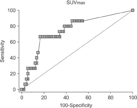
Fig. 1 Receiver operating characteristic curve of maximum standardized uptake valvue (SUVmax) for predicting underestimation of invasive breast cancer. Optimal cutoff of SUVmax was 2.65 (n = 102).
Reference
-
1. Min SY, Kim Z, Hur MH, Yoon CS, Park EH, Jung KW, et al. The basic facts of Korean breast cancer in 2013: results of a Nationwide Survey and Breast Cancer Registry Database. J Breast Cancer. 2016; 19:1–7.
Article2. Siziopikou KP. Ductal carcinoma in situ of the breast: current concepts and future directions. Arch Pathol Lab Med. 2013; 137:462–466.
Article3. Park AY, Gweon HM, Son EJ, Yoo M, Kim JA, Youk JH. Ductal carcinoma in situ diagnosed at US-guided 14-gauge core-needle biopsy for breast mass: preoperative predictors of invasive breast cancer. Eur J Radiol. 2014; 83:654–659.
Article4. Brennan ME, Turner RM, Ciatto S, Marinovich ML, French JR, Macaskill P, et al. Ductal carcinoma in situ at core-needle biopsy: meta-analysis of underestimation and predictors of invasive breast cancer. Radiology. 2011; 260:119–128.
Article5. Ueda S, Tsuda H, Asakawa H, Shigekawa T, Fukatsu K, Kondo N, et al. Clinicopathological and prognostic relevance of uptake level using 18F-fluorodeoxyglucose positron emission tomography/computed tomography fusion imaging (18F-FDG PET/CT) in primary breast cancer. Jpn J Clin Oncol. 2008; 38:250–258.
Article6. Kadoya T, Aogi K, Kiyoto S, Masumoto N, Sugawara Y, Okada M. Role of maximum standardized uptake value in fluorodeoxyglucose positron emission tomography/computed tomography predicts malignancy grade and prognosis of operable breast cancer: a multi-institute study. Breast Cancer Res Treat. 2013; 141:269–275.
Article7. Chin-Lenn L, Mack LA, Temple W, Cherniak W, Quinn RR, Ravani P, et al. Predictors of treatment with mastectomy, use of sentinel lymph node biopsy and upstaging to invasive cancer in patients diagnosed with breast ductal carcinoma in situ (DCIS) on core biopsy. Ann Surg Oncol. 2014; 21:66–73.
Article8. Sato Y, Kinoshita T, Suzuki J, Jimbo K, Asaga S, Hojo T, et al. Preoperatively diagnosed ductal carcinoma in situ: risk prediction of invasion and effects on axillary management. Breast Cancer. 2016; 23:761–770.
Article9. Park AY, Gweon HM, Son EJ, Yoo M, Kim JA, Youk JH. Ductal carcinoma in situ diagnosed at US-guided 14-gauge core-needle biopsy for breast mass: preoperative predictors of invasive breast cancer. Eur J Radiol. 2014; 83:654–659.
Article10. Park SH, Kim MJ, Kim SJ, Kim EK. Ductal carcinoma in situ diagnosed using an ultrasound-guided 14-gauge core needle biopsy of breast masses: can underestimation be predicted preoperatively? Ultrasonography. 2014; 33:128–135.
Article11. Shin SH, Kim BC, Song YJ, Yoon HC, Cho JS, Park MH, et al. Risk factor of invasive breast cancer in patients with preoperative diagnosis of ductal carcinoma in situ. J Korean Surg Soc. 2011; 80:90–95.
Article12. Nori J, Meattini I, Giannotti E, Abdulcadir D, Mariscotti G, Calabrese M, et al. Role of preoperative breast MRI in ductal carcinoma in situ for prediction of the presence and assessment of the extent of occult invasive component. Breast J. 2014; 20:243–248.13. Berriolo-Riedinger A, Touzery C, Riedinger JM, Toubeau M, Coudert B, Arnould L, et al. [18F]FDG-PET predicts complete pathological response of breast cancer to neoadjuvant chemotherapy. Eur J Nucl Med Mol Imaging. 2007; 34:1915–1924.
Article14. Ohara M, Shigematsu H, Tsutani Y, Emi A, Masumoto N, Ozaki S, et al. Role of FDG-PET/CT in evaluating surgical outcomes of operable breast cancer: usefulness for malignant grade of triple-negative breast cancer. Breast. 2013; 22:958–963.15. Morris PG, Ulaner GA, Eaton A, Fazio M, Jhaveri K, Patil S, et al. Standardized uptake value by positron emission tomography/computed tomography as a prognostic variable in metastatic breast cancer. Cancer. 2012; 118:5454–5462.
Article16. Shigematsu H, Kadoya T, Masumoto N, Matsuura K, Emi A, Kajitani K, et al. Role of FDG-PET/CT in prediction of underestimation of invasive breast cancer in cases of ductal carcinoma in situ diagnosed at needle biopsy. Clin Breast Cancer. 2014; 14:358–364.17. Hashimoto Y, Tsujikawa T, Kondo C, Maki M, Momose M, Nagai A, et al. Accuracy of PET for diagnosis of solid pulmonary lesions with 18F-FDG uptake below the standardized uptake value of 2.5. J Nucl Med. 2006; 47:426–431.18. Herrmann K, Czernin J, Cloughesy T, Lai A, Pomykala KL, Benz MR, et al. Comparison of visual and semiquantitative analysis of 18F-FDOPA-PET/CT for recurrence detection in glioblastoma patients. Neuro Oncol. 2014; 16:603–609.
Article
- Full Text Links
-
- Actions
-
Cited
- CITED
-
- Close
- Share
-
- Similar articles
-
- 18F-Fluorodeoxyglucose Positron Emission Tomography/CT Scan Findings for Ductal Carcinomas of Breast: Association of Standardized Uptake Value and Histological Findings
- Unusual Horner's Syndrome in Recurrent Breast Cancer: Evaluation Using ¹â¸F-FDG PET/CT
- Effectiveness of Breast MRI and 18F-FDG PET/CT for the Preoperative Staging of Invasive Lobular Carcinoma versus Ductal Carcinoma
- Invasive Lobular Carcinoma of the Breast Associated with Mixed Lobular and Ductal Carcinoma In Situ: A Case Report
- Dual Pathologies of Parathyroid Adenoma and Papillary Thyroid Cancer on Fluorocholine and Fluorodeoxyglucose PET/CT
