Expression of Erythropoietin in the Spinal Cord of Lewis Rats with Experimental Autoimmune Encephalomyelitis
- Affiliations
-
- 1Department of Neurology, College of Medicine, Jeju National University, Jeju, Korea.
- 2Department of Neurosurgery, College of Medicine, Jeju National University, Jeju, Korea.
- 3Department of Veterinary Medicine, College of Medicine, Jeju National University, Jeju, Korea. shint@cheju.ac.kr
Abstract
-
BACKGROUND AND PURPOSE: Erythropoietin (Epo), originally recognized for its central role in erythropoiesis, has been shown to improve the outcomes in patients with various neurological disorders. The aim of this study was to elucidate the Epo expression pattern in the spinal cords of Lewis rats with experimental autoimmune encephalomyelitis (EAE) and to assess the systemic effect of Epo during the course of EAE.
METHODS
We used an EAE model induced in Lewis rats by immunization with myelin basic protein. Immunized rats were given recombinant human Epo (rhEpo) intraperitoneally at a dose of 5,000 U/kg for 7 consecutive days, either starting on day 3 post-immunization (five rats) or on the day of clinical symptom onset (score > or =1, five rats). After immunization, the rats were observed daily for clinical signs of EAE. Epo expression was investigated by Western blot analysis and immunohistochemistry.
RESULTS
Western blot analysis showed that, Epo expression was significantly elevated relative to control in the rat spinal cord during the peak stage of EAE (p<0.05), and then decreased thereafter. Immunohistochemistry demonstrated that Epo was expressed in some neurons and glial cells. Epo immunoreactivity was detected in ED1-positive macrophages and astrocytes in EAE lesions. Furthermore, we found that the intraperitoneal administration of rhEpo reduced both the disease severity and duration of paralysis in EAE rats, and reduced macrophage activity and increased Epo activity.
CONCLUSIONS
Based on these findings, we postulate that Epo expression begins to increase at the start of EAE and that rhEpo administration leads to functional recovery from EAE paralysis.
MeSH Terms
Figure
-

Fig. 1 Western blot analysis of Epo expression in the spinal cord of rats with EAE. Spinal cord samples were collected from normal control rats, rats at the peak stage of EAE (G.3, day 12 PI), and rats during the recovery stage of EAE (R.0, day 21 PI). Representative photographs of Western blots; arrowheads indicate the expression of Epo and β-actin (upper). Semiquantitative analysis of Epo immunoreactivity in spinal cords, normalized to the intensity of β-actin expression in the same immunoblot. Epo was detected in the normal controls, and its expression was significantly increased in EAE-affected spinal cords. Data are (mean±SEM values) from three experiments (lower), *p<0.05 vs. normal controls and the recovery stage of EAE, †p<0.05 vs. normal controls. PI: post-immunization, Epo: erythropoietin, EAE: experimental autoimmune encephalomyelitis.
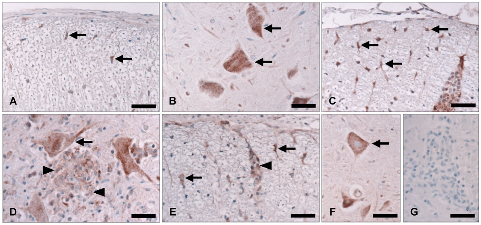
Fig. 2 Immunohistochemical staining of Epo in the spinal cords of normal control rats (A and B) and rats at the peak (day 12 PI)(C and D) and recovery (day 21 PI)(E and F) stages of EAE. Epo was weakly detected in some glial cells (A, arrows) and neurons (B, arrows) in the spinal cords of normal control rats. Glial cells at the peak stage of EAE showed increased immunoreactivity for Epo (C, arrows). Epo-positive inflammatory cells (D, arrowheads) and neurons (D, arrow) were detected in the parenchyma. At the recovery stage of EAE, some inflammatory cells (E, arrowhead), glial cells (E, arrows) and neurons (F, arrows) were positive for Epo. Tissue from a rat at the peak stage of EAE stained without the primary antisera showed no staining (G). Counterstaining was with hematoxylin. Scale bars=40 µm. Epo: Erythropoietin, PI: post-immunization, EAE: experimental autoimmune encephalomyelitis.
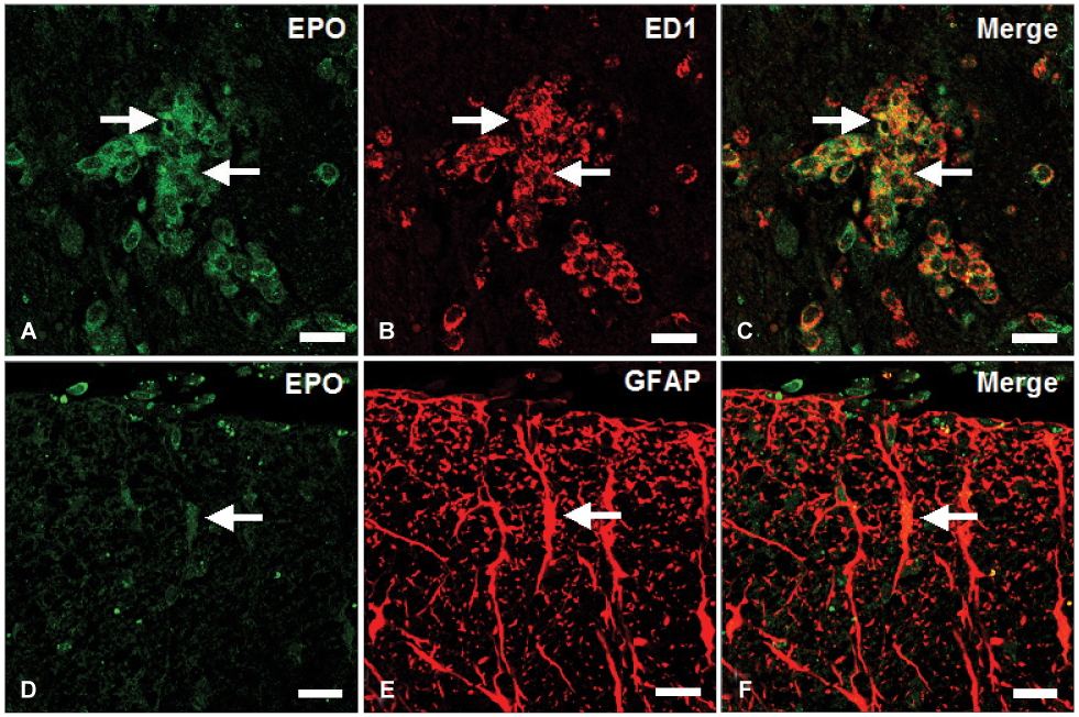
Fig. 3 Immunofluorescent co-localization of Epo (A and D) with ED1 (B) and anti-GFAP (E) in the spinal cords of rats at the peak stage of EAE (day 12 PI). A large amount of Epo (A, green; arrows) was immunostained in ED1-positive macrophages (B, red; arrows)(C, merge; arrows). Epo immunoreactivity (D, green; arrow) was co-localized in GFAP (E, red; arrow)-positive astrocytes (F, merge; arrow). Scale bars: A-F, 20 µm. Epo: Erythropoietin, anti-GFAP: anti-glial fibrillary acidic protein, EAE: experimental autoimmune encephalomyelitis, PI: post-immunization.
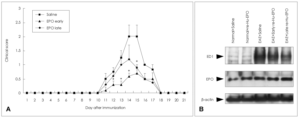
Fig. 4 Effect of recombinant human erythropoietin (rhEpo) therapy on the clinical course and protein expression in EAE. A: Rats were immunized with myelin basic protein and injected with saline (vehicle), rhEpo (5,000 U/kg) from day 3 PI (early group), or rhEpo from the day of clinical symptom onset (late group). Rats were scored daily for clinical signs of EAE. Data are the mean±SEM values from five animals per group. B: Western blot analysis of ED1 and Epo expressions in the Epo-treatment experiment. Spinal cord samples were collected from normal rats that were injected with saline (lane 1), rhEpo (lane 2), and from EAE-affected rats (day 15 PI) that were injected with vehicle (lane 3), rhEpo from day 3 PI (lane 4), and Epo from the day of clinical symptom onset (lane 5). *p<0.05 vs. vehicle. EAE: experimental autoimmune encephalomyelitis, PI: post-immunization.
Reference
-
1. Sirén AL, Ehrenreich H. Erythropoietin--a novel concept for neuroprotection. Eur Arch Psychiatry Clin Neurosci. 2001. 251:179–184.2. Buemi M, Cavallaro E, Floccari F, Sturiale A, Aloisi C, Trimarchi M, et al. The pleiotropic effects of erythropoietin in the central nervous system. J Neuropathol Exp Neurol. 2003. 62:228–236.
Article3. Brines M, Cerami A. Emerging biological roles for erythropoietin in the nervous system. Nat Rev Neurosci. 2005. 6:484–494.
Article4. Marti HH. Erythropoietin and the hypoxic brain. J Exp Biol. 2004. 207:3233–3242.
Article5. Shingo T, Sorokan ST, Shimazaki T, Weiss S. Erythropoietin regulates the in vitro and in vivo production of neuronal progenitors by mammalian forebrain neural stem cells. J Neurosci. 2001. 21:9733–9743.
Article6. Yu X, Shacka JJ, Eells JB, Suarez-Quian C, Przygodzki RM, Beleslin-Cokic B, et al. Erythropoietin receptor signaling is required for normal brain development. Development. 2002. 129:505–516.
Article7. Brines ML, Ghezzi P, Keenan S, Agnello D, de Lanerolle NC, Cerami C, et al. Erythropoietin crosses the blood-brain barrier to protect against experimental brain injury. Proc Natl Acad Sci U S A. 2000. 97:10526–10531.
Article8. Chung YH, Kim SI, Joo KM, Kim YS, Lee WB, Yun KW, et al. Age-related changes in erythropoietin immunoreactivity in the cerebral cortex and hippocampus of rats. Brain Res. 2004. 1018:141–146.
Article9. Wang L, Zhang Z, Wang Y, Zhang R, Chopp M. Treatment of stroke with erythropoietin enhances neurogenesis and angiogenesis and improves neurological function in rats. Stroke. 2004. 35:1732–1737.
Article10. Chung YH, Joo KM, Kim YS, Lee KH, Lee WB, Cha CI. Enhanced expression of erythropoietin in the central nervous system of SOD1 (G93A) transgenic mice. Brain Res. 2004. 1016:272–280.
Article11. Agnello D, Bigini P, Villa P, Mennini T, Cerami A, Brines ML, et al. Erythropoietin exerts an anti-inflammatory effect on the CNS in a model of experimental autoimmune encephalomyelitis. Brain Res. 2002. 952:128–134.
Article12. Zhang J, Li Y, Cui Y, Chen J, Lu M, Elias SB, et al. Erythropoietin treatment improves neurological functional recovery in EAE mice. Brain Res. 2005. 1034:34–39.
Article13. Li W, Maeda Y, Yuan RR, Elkabes S, Cook S, Dowling P. Beneficial effect of erythropoietin on experimental allergic encephalomyelitis. Ann Neurol. 2004. 56:767–777.
Article14. Savino C, Pedotti R, Baggi F, Ubiali F, Gallo B, Nava S, et al. Delayed administration of erythropoietin and its non-erythropoietic derivatives ameliorates chronic murine autoimmune encephalomyelitis. J Neuroimmunol. 2006. 172:27–37.
Article15. Ruscher K, Freyer D, Karsch M, Isaev N, Megow D, Sawitzki B, et al. Erythropoietin is a paracrine mediator of ischemic tolerance in the brain: evidence from an in vitro model. J Neurosci. 2002. 22:10291–10301.
Article16. Sirén AL, Knerlich F, Poser W, Gleiter CH, Brück W, Ehrenreich H. Erythropoietin and erythropoietin receptor in human ischemic/hypoxic brain. Acta Neuropathol. 2001. 101:271–276.
Article17. Rotshenker S. Microglia and macrophage activation and the regulation of complement-receptor-3 (CR3/MAC-1)-mediated myelin phagocytosis in injury and disease. J Mol Neurosci. 2003. 21:65–72.
Article18. Hammarberg H, Lidman O, Lundberg C, Eltayeb SY, Gielen AW, Muhallab S. Neuroprotection by encephalomyelitis: rescue of mechanically injured neurons and neurotrophin production by CNS-infiltrating T and natural killer cells. J Neurosci. 2000. 20:5283–5291.
Article19. Trzonkowski P, Debska-Slizien A, Szmit E, Myśliwska J, Szymańska K, Hak Ł, et al. Long-term therapy with recombinant human erythropoietin increases CD8+ T-cell apoptosis in haemodialysis patients. Nephrol Dial Transplant. 2005. 20:367–376.
Article20. Shin T, Kojima T, Tanuma N, Ishihara Y, Matsumoto Y. The subarachnoid space as a site for precursor T cell proliferation and effector T cell selection in experimental autoimmune encephalomyelitis. J Neuroimmunol. 1995. 56:171–178.
Article21. Moon C, Kim S, Wie M, Kim H, Cheong J, Park J, et al. Increased expression of p53 and Bax in the spinal cords of rats with experimental autoimmune encephalomyelitis. Neurosci Lett. 2000. 289:41–44.
Article
- Full Text Links
-
- Actions
-
Cited
- CITED
-
- Close
- Share
-
- Similar articles
-
- Increased expression of neuronal nitric oxide synthase in astrocytes and macrophages in the spinal cord of Lewis rats with autoimmune encephalomyelitis
- Erythropoietin and autoimmune neuroinflammation: lessons from experimental autoimmune encephalomyelitis and experimental autoimmune neuritis
- Melatonin ameliorates autoimmune encephalomyelitis through suppression of intercellular adhesion molecule-1
- Increased expression of osteopontin in the spinal cords of Lewis rats with experimental autoimmune neuritis
- Immunohistochemical Localization of Bcl-2 in the Spinal Cords of Rats with Experimental Autoimmune Encephalomyelitis
